About The Lab
A hallmark of the Alzheimer disease brain is the presence of the intracellular neurofibrillary tangles composed primarily of the protein tau in a pathologically modified state. There is compelling evidence that the accumulation of tau with aberrant posttranslational modifications is central to the disease process and that the abnormally modified tau is toxic to neurons. This increased accumulation of tau is likely due in part to inefficient clearance mechanisms. Therefore an understanding of the proteolytic processes that degrade and remove tau from neurons is needed. In this context a primary focus of our lab is on delineating the mechanisms involved in clearing tau from neurons. Based on previous studies it is clear that autophagy is a primary degradative pathway in neurons. We and others have provided evidence that tau is degraded by autophagy, but the processes involved have not been clearly identified. Currently ongoing research in the lab is directed towards understanding how tau is selectively targeted to autophagy for degradation and how these processes could be dysregulated in Alzheimer disease.
Another area of ongoing investigation, in collaboration with the Nehrke lab, is on understanding the mechanisms by which Alzheimer’s disease relevant forms of tau cause neuron toxicity. For these studies we use an invertebrate animal model; C. elegans that express single copies of different forms of potentially pathological tau in specific neuron populations. We then are able to assess the effects of tau on neuron morphology and function, axonal transport and mitochondrial function, among other possible targets. Results from these studies will then be transferred into mammalian model systems.
The Johnson lab also has a well-established interest in understanding the regulation and function of transglutaminase 2 (TG2) in neural cell death and survival, particular in the context of central nervous system injury. We have found that in neurons TG2 promotes survival subsequent to ischemic injury and this is dependent on TG2 localization to the nucleus. Interestingly, TG2 in astrocytes has the opposite effect as it plays a detrimental role in injury responses. Deletion or inhibition of TG2 from astrocytes significantly increases their ability to survive stress-induced cell death and to protect neurons from oxygen and glucose deprivation-induced cell death.
Additionally, promising data suggest that specific deletion of TG2 from astrocytes improves outcomes after spinal cord injury. Future studies will involve testing TG2 inhibitors as therapeutics for the treatment of spinal cord injuries.
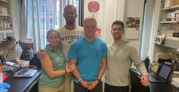
In addition to the exciting research, our group has fun together our side the lab! We recently had fun as a group ziplining! No doubt there will be more thrilling adventures in the future!
