Microscopy with UV Surface Excitation (MUSE) Imaging
Microscopic evaluation of tissue pathology is typically divided between intraoperative techniques such as frozen sections analysis, which is expensive and only available in limited scenarios, and conventional paraffin embedded histology, which is available to most physicians, but requires several days delay for tissue processing. MUSE imaging is an emerging technique that rapidly (seconds) produces histological images using ultraviolet LEDs to excite fluorescence from the surface layers of whole tissue specimens. MUSE can be combined with virtual transmission microscopy algorithms to produce images similar to conventional histology, or can be used to extract novel sources of contrast.
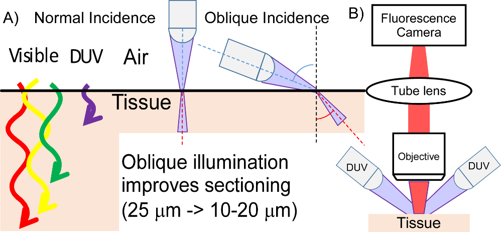
Deep ultraviolet (DUV) imaging with MUSE. DUV light selectively excites the surface of tissue, enabling inexpensive imaging of pathology.
A key advantage of MUSE is the very low system cost and complexity, which enables application of rapid histology in areas for which is it currently not cost-effective. In select applications, MUSE can provide similar image contrast as existing methods, but at greatly reduced processing time and cost.
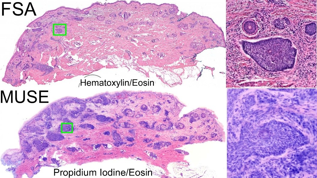
Imaging of a skin cancer specimen using conventional frozen sections analysis (FSA) and with low-cost MUSE imaging. MUSE shows similar details to FSA but is dramatically faster and less expensive.