Multiphoton Skin Biopsy Imaging
Nonmelanoma skin cancer (NMSC) stands as the most prevalent form of cancer in the United States, surpassing the combined annual cases of other cancers. The standard diagnostic approach for NMSC is formalin-fixed paraffin-embedded (FFPE) processes with hematoxylin and eosin (H&E) staining. This process, however, necessitates a waiting period of several days before patients receive their diagnostic results, subsequently delaying surgical scheduling and adding to patient inconvenience.
Recent advancements have demonstrated the efficacy of compact multiphoton microscopes with virtual H&E rendering for histological analysis, in delivering rapid tissue imaging. This innovative approach promises the possibility of same-day biopsy diagnoses, significantly curtailing the waiting period. Consequently, it facilitates same-day treatment, enhancing clinical efficiency and patient convenience. Our latest pilot study demonstrated this potential, indicating that our advanced compact multiphoton system can provide rapid, and effective point-of-care diagnoses for NMSC.
Current ongoing clinical studies are focusing on the implementation of modified specimen holders. These modifications enable similar cross-sectional views as FFPE images and improving good co-registration with FFPE images.
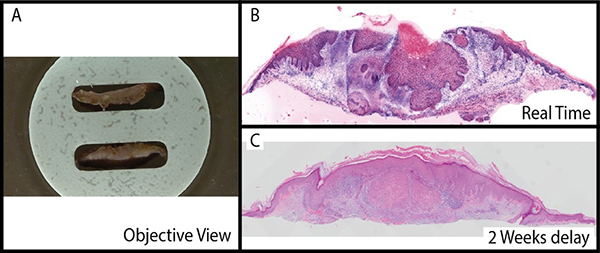
Figure 1: Shave skin biopsy from a 74-year-old white, non-Hispanic male presenting with a suspicious lesion on the left upper forearm. The patient consented and was imaged minutes after the shave biopsy, revealing squamous cell carcinoma (top). Two weeks later, conventional paraffin sections confirmed the diagnosis (bottom).