News
Payea and Mishra are Inaugural Recipients of the Sayeeda Zain Travel Award
Friday, July 14, 2017
The department of Biochemistry and Biophysics recently presented to the inaugural Sayeeda Zain Travel Award to Mathew Payea and Laxmi Narayan Mishra.
Matthew Payea is a 6th year graduate student in the Biochemistry Ph.D. program studying tRNA biology in laboratory of Eric Phizicky. Matthew received his Bachelors in Science from Eastern Illinois University, majoring in Biochemistry. Matthew used the funding provided by the Sayeeda Zain Travel Award to attend the 22nd annual meeting of the RNA Society in Prague, Czech Republic this past June. There, he gave a talk on his research defining an RNA decay pathway in yeast that destroys mutant tRNAs.
Laxmi Narayan Mishra is a postdoctoral associate working in Dr. Jeffrey J Hayes' Lab in the Department of Biochemistry and Biophysics, University of Rochester Medical Center. He has a Masters degree from University of North Bengal, Darjeeling, India and a Ph.D. in Biochemistry from Jawaharlal Nehru Centre for Advanced Scientific Research, Bangalore, India. His research is focused on how epigenetic modifications alter chromosome structure to facilitate gene expression. His Dr. Mishra will use the Sayeeda Zain Travel Award to attend and present his research findings at the international EMBO conference on "The Nucleosomes: From Atoms to Genomes" at Heidelberg, Germany, in August 2017.
The Sayeeda Zain Travel Award is given semi-annually to one or more graduate students and postdoctoral fellows in the Department of Biochemistry and Biophysics at the University of Rochester School of Medicine and Dentistry. The award honors the life and achievements of Professor Sayeed Zain, Ph.D., a longtime faculty member in the Department of Biochemistry and Biophysics. Learn more about the award and Dr. Zain.
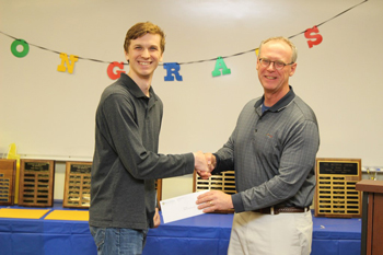
Matthew Payea (left) with Dr. Jeffrey Hayes
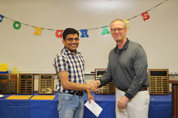
Laxmi Narayan Mishra (left) with Dr. Jeffrey Hayes
Jeffrey Hayes Helps Increase Our Understanding of DNA's Path Into Cells
Thursday, June 29, 2017
Scientists are a step closer to understanding how DNA, the molecules that carry all of our genetic information, is squeezed into every cell in the body. How DNA is "packaged" in cells influences the activity of our genes and our risk for disease. Elucidating this process will help researchers in all areas of health care, from cancer and heart disease, to muscular dystrophy and osteoarthritis.
DNA is a long, floppy molecule, and there's more than three feet of it in every cell. Our DNA is housed in structures called chromosomes, which condense the DNA to fit into the cell's tight quarters.
Scientists from the department of Biochemistry and Biophysics at the University of Rochester School of Medicine and Dentistry worked with colleagues in France and Japan to describe the first step of DNA packing in a cell. They provided the first-ever detailed picture of the most basic building block of chromosomes, known as the nucleosome, and found that a protein known as H1 (for linker histone H1) helps DNA become more compact and rigid within the nucleosome. In contrast, when H1 isn't present, the DNA is loose and flexible.
The tight structure that H1 creates helps shield our DNA from various factors that can activate or "turn on" certain genes. Without H1, DNA is more accessible to factors that could trigger disease-causing genes.
Published in the journal Molecular Cell, this finding will inform research on all processes that involve chromosomes, such as gene expression and DNA repair, which are critical to the understanding of diseases such as cancer, according to Jeffrey J. Hayes, Ph.D., senior study author and the Shohei Koide Professor and chair of the department of Biochemistry and Biophysics.
The teams in France and Japan used specialized microscopes and X-rays to capture pictures of DNA molecules interacting with H1 and other key proteins. Because of the size of the DNA and protein molecules, the pictures generated by these techniques were fuzzy and difficult to analyze.
Lead study author Amber Cutter, a graduate student in Hayes' lab, put all of the components -- DNA, H1, and other proteins -- together in tiny test tubes and conducted various biochemical experiments. Her tests, coupled with the X-ray images, confirmed H1's role.
Cutter (pictured at right), who is entering her fifth year in Hayes' lab, admits that the science is complex and that a lot more research needs to be done before this work can inform clinical treatment. But, the importance of understanding the most basic biological processes should not be underestimated. "In order to determine what happens when things go wrong in diseases like cancer, we need to know what happens when things go right."
Hayes and Cutters' work was supported by the National Institutes of Health.
# # #
The University of Rochester Medical Center is home to approximately 3,000 individuals who conduct research on everything from cancer and heart disease to Parkinson's, pandemic influenza and autism. Spread across many centers, institutes and labs, our scientists have developed therapies that have improved human health locally, in the region and across the globe. To learn more, visit www.urmc.rochester.edu/research.