RF Coil Development
RF Coil Development
RF Coil
We have been developing specially designed RF coils for both research and clinical applications at 1.5T and 3T. Through new coil design concepts and optimal design for each individual application, these coils improve image signal-to-noise ratio (SNR), and thereby increase image resolution and reduce scan time.
Monkey brain phased array coil
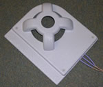
A dedicated receiver coil for monkey brain MR imaging was developed for use at 3T. It is based on a new three-dimensional orthogonal phased array design that is first reported. In vivo results show that this coil provides high and uniform signal to support high-resolution imaging of the whole monkey brain (a,b). Moreover, it is compatible with parallel imaging that reduces susceptibility-induced image distortion in EPI scans (c-f). It is useful for monkey brain modeling studies of diseases, brain development and brain functions.
Figure A. High
resolution image of
monkey brain.
Figure B. High
resolution image of
monkey brain.
Figure C. EPI scan.
Figure D. EPI scan.
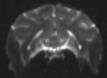
Figure E. EPI scan.
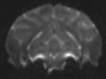
Figure F. EPI scan.
Liquid-nitrogen cooled phased array coil
A liquid-nitrogen cooled (LN2) dual-channel array coil was developed for use at 3T (a). Each of the coil elements is 3.5cm x 3.5cm in size, and the two elements are partially overlapped to eliminate coupling between them. The image data of human fingers (b) and mouse (c) show that the LN2 coil increases SNR by more than 100% compared with a similar coil at room temperature. With this SNR increase, image resolution may be doubled or scan time may be reduced by a factor of four.
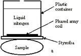
Figure A. Liquid-nitrogen
cooled (LN2) dual-channel
array coil diagram.

Figure B. Image of
human fingers.
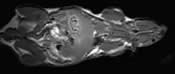
Figure C. Image of a mouse.
Selected Publications/Presentations
- Kwok WE, You Z, Monu J, Seo G, Ritchlin C. High-Resolution Uniform MR Imaging of Finger Joints Using a Dedicated RF Coil at 3 Tesla. J Magn Reson Imaging 2010;31:240-247.
- You Z, Kwok WE, Proulx S, Schwarz EM. Simultaneous High-Resolution Imaging of Mouse Knee and Ankle at 3.0T Using a Specially Designed Dual Array Coil. In: Proceedings of ISMRM 16th Scientific Meeting and Exhibition, Toronto, Canada 2008, p3644.
- You Z, Kwok WE, Mukherjee M, Mancarella M. 3D-Orthogonal Phased Array Coil for High-Resolution and Low-distortion EPI Imaging of Monkey Brain at 3.0T. In: Proceedings of ISMRM 15th Scientific Meeting and Exhibition, Berlin, Germany 2007, p 1040.
- Kwok WE, You Z. In vivo MR imaging using liquid nitrogen cooled phased array coil at 3.0 Tesla. Magn Reson Imaging 2006;24:819-823.
- Kwok WE, Zhong J, You Z, Seo GS, Totterman SMS. A Four-element phased array coil for high resolution and parallel MR imaging of the knee. Magn Reson Imaging 2003;21:961-967.
- Kwok WE, Lo KK, Seo G, Totterman SMS. A volume adjustable four-coil phased array for high resolution MR imaging of the hip. Magnetic Resonance Materials in Physics, Biology & Medicine 1999; 9(1/2):59-64.
Invited Presentations
- “The Development of Specialized Coils for Musculoskeletal Imaging” presented at the Musculoskeletal MR Study Group at the 14th Annual Meeting of the International Society of Magnetic Resonance in Medicine, Seattle, WA on 5/2006.
- “Specialized MRI Coils for Research and Clinical Applications” presented at Tokyo Dental College, Chiba, Japan on 4/2007.
- “Specialized RF coils for human and animal MR imaging at 3T” presented at the University of Hong Kong, Hong Kong on 4/2007.