Multi-scaled Computational Analysis of Cochlear Mechano-transduction
Multi-scaled Computational Analysis of Cochlear Mechano-transduction
Project Collaborator:
The mammalian cochlea is a mechano-transducer—it turns acoustic energy into electro-chemical energy. The cochlea not only receives external stimuli, but emits sound. These emissions are referred to as reverse transduction to distinguish from the acoustic to electric (forward) transduction. The reverse transduction is a hallmark of a sensitive (functional) cochlea. The reverse transduction (otoacoustic emissions) has been extensively measured and analyzed. However, the source and the generation mechanism of the reverse transduction remain poorly understood.

Multi-scaled model of cochlear mechano-transduction
(See enlargement)
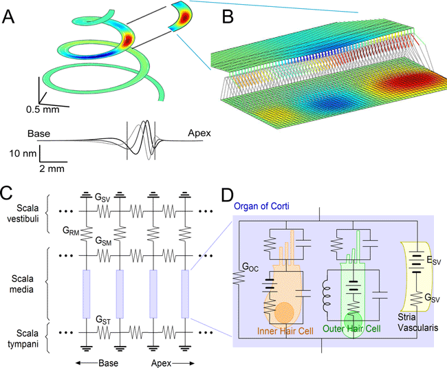
The image to the right is a multi-scaled model of cochlear mechano-transduction. (A) Fluid-structure interaction of the cochlea coil. At 1 kHz sound stimulus, the wave travels to 8 mm from basal end before peaking. (B) A 900 µm piece of the partition will be analyzed in further detail using finite element method that includes active electro-mechanical OHCs. (C) Electrical representation of the cochlear duct. Three transmission lines along three ducts (scala vestibule, media and tympani). (D) The OC will be further detailed. It has circuits of IHC, OHC and stria vascularis. All other conductance across the epithelium is lumped to GOC. These mechanical and electrical models will be analyzed simultaneously.
In order to better understand the cochlear mechano-transduction, we develop a multi-scale computational model that incorporates the kinetics of the mechano-transduction channels, the mechanics of the hair-bundle, the electrical circuit of hair cells including their motility, the fluid mechanics of IHC, the electro-mechanics of the organ of Corti and the entire cochlea. Specifically we are investigating the mechanics of the organ of Corti, the sensory epithelium in the cochlea. Full 3-D mechanics of the organ of Corti are analyzed in the time domain (see movie clip below). The emphasis will be on
- The role of the tectorial membrane
- The active force transmission from the outer hair cells to the basilar membrane.
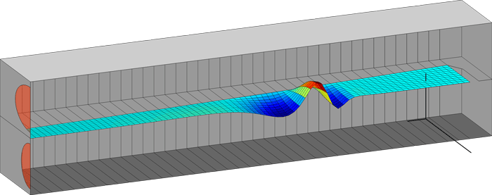
Computer simulation of cochlear fluid mechanics
Above is a computer simulation of cochlear fluid mechanics. The cochlea was modeled as a rectangular box filled with lymphatic fluid and divided by an elastic partition. As the oval window (the upper red circle in the left) vibrates the cochlear fluid is pressurized. The pressurized fluid creates traveling displacement waves along the partition. The model is 12.5 mm long and 0.5 mm wide. (Yanju Liu, ME PhD student).
Because no commercial program provides an easier way to solve these multi-scale and multi-physics problems, we develop our own program in Matlab. Computer codes of our published works are available upon request.