URMC / Labs / Topham Lab / Projects / Dynamic regulation of CD8+ T cell motility in the influenza infected airway
Dynamic regulation of CD8+ T cell motility in the influenza infected airway
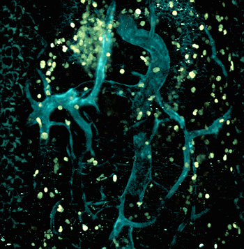
Image of an influenza infected trachea obtained by multiphoton microscopy. This image shows blood vessels and epithelial cells in blue, and CD8 T cells in green. The T cells are the immune effectors responsible for killing virus infected cells in the airways.
CD8+ T cells can control influenza infection by killing infected cells so they can no longer make virus. Because influenza only infects the cells that line the respiratory tract, they must be able to travel through the circulation, exit the vasculature, and migrate within the airway tissues to locate the virus infected cells. This is a complex, multistep process. T cells interact with the tissue through various receptors that include integrin adhesion molecules that bind extracellular matrix and other cells, and chemokines that guide the T cells towards areas of infection and to antigen presenting cells in the tissue. We study how the tissue is modified during the infection and the resulting immune response, as well as how the T cells adapt and respond to these modifications. To perform these studies on cell motility and migration, we have developed sophisticated genetic and fluorescent reporters for the T cells, the virus, and non-immune cells in the tissue combined with a multicolor live imaging platform that can collect information on the cells in real time. We use these tools to study the CD8 T cells during acute infection, in the formation of tissue-resident memory (TRM), and in reactivation of the cells after secondary infection. Our goals are to better understand the regulation mechanisms of the T cells and tissue micro-environment, which will lead to novel strategies to improve immune protection or modulate immune pathogenesis and disease severity.