Stroke
Make Appointments & Get Care
Time is Brain. Suspect a Stroke? Call 911.
If you have any sudden or unexplained face drooping, arm weakness, or speech difficulty, call 911 immediately. Every second counts.
What is a Stroke?
A stroke is a medical emergency where every second counts. A stroke results from the blockage (ischemic stroke) or bursting (hemorrhagic stroke) of blood vessels to the brain. This means not enough oxygen reaches brain tissue, so brain cells begin to die. As this happens, patients experience loss of function/malfunction of that region of the brain.
About one quarter of patients experience a warning sign—a mini-stroke (called a transient ischemic attack, or TIA). A TIA does not leave permanent damage but warns that a more serious stroke is coming soon. It’s important that TIAs are treated urgently to prevent future strokes.
University of Rochester Medicine offers a Rapid Access TIA Clinic as well emergency telestroke consultations via video technology to swiftly address these events.
URochester Medicine is home to the following types of stroke centers:
- The Comprehensive Stroke Center at Strong Memorial Hospital is equipped to handle the most complex of cases.
- Our Primary Stroke Centers at Highland Hospital, Thompson Health, Noyes Memorial Hospital, Geneva General Hospital, and Jones Memorial Hospital.
We also offer the region’s only Mobile Stroke Unit, which brings the resources of an emergency room directly to the stroke patient’s driveway.
Remember the phrase FAST or BE FAST:
Balance: loss of balance
Eyes: vision issues
Face: is one side drooping?
Arms: is one arm hard to lift?
Speech: is speech slurred or confused?
Time: if you see any of those signs, it’s time to call 911 immediately. Every second counts.
Stroke in Our Community
Stroke happens more often and is more dangerous in parts of our community, especially for Black and Hispanic adults. Knowing the signs and how to prevent stroke are key.
Life Saving Care
URochester Medicine Mobile Stroke Unit is the region’s only ambulance equipped to treat strokes. It has a CT scanner on board to diagnose and start treatment before reaching the hospital.
STAR-NY
The Stroke Treatment Alliance of New York works to improve stroke care through collaboration and shared best practices. It brings health systems together to provide high-quality treatment for patients.
Treatments for Stroke
How is a Stroke Diagnosed?
Stroke patients are assessed quickly in the emergency department by a specialized stroke team that includes vascular neurologists, neurosurgeons, and emergency physicians.
A variety of imaging techniques are used in stroke diagnosis, including:
By these techniques the doctors can pinpoint the area of stroke in the brain as well as the area of blockage of the vessels.
In patients with an “ischemic stroke” (i.e., no blood is seen on their initial CT scan) who arrive in time, a clot-busting medication called tPA can be used to open blocked arteries.
In patients with hemorrhagic stroke (bleeding in the brain), control of blood-clotting factors, blood pressure, and even emergency surgery might be needed.
After initial treatment, patients are either admitted to a dedicated stroke unit or may first require critical care in our neurointensive care unit.
Surgical Treatments for Stroke
Some patients need emergency surgery to treat blocked or bleeding brain blood vessels.
Our neurosurgeons are fellowship-trained to use special catheters (tiny tubes) and devices to open blocked brain arteries to restore blood flow to the brain.
Following recovery from stroke, our cerebrovascular neurosurgeons can perform surgery or stenting to open a narrowed artery that caused the initial stroke, helping prevent future problems.
Patients with stroke caused by leaking blood vessels (such as burst brain aneurysms or AVM) may require embolization, which uses wires and catheters inside the arteries, and/or open brain surgery.

Can You Have a Stroke and Not Know It?
Learn the signs of silent stroke, early stroke symptoms, differences between men and women, and what to do in an emergency.
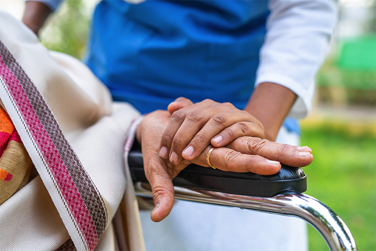
Stroke Recovery & Rehabilitation
Our multidisciplinary stroke team helps manage all aspects of recovery.
While some patients recover within days, it is not unusual for recovery to take weeks to months.
To help stroke patients recover to their maximum potential, each patient is evaluated for their physical, occupational, and speech therapy needs, along with intensive rehabilitation and preventative care to minimize chances of a future stroke.
Neurological rehabilitation may consist of physical or occupational therapy, medicine prescription, modalities, orthotics / bracing, speech and language therapy, or neuropsychology and referrals to other disciplines when needed.
Patient Stories
Kathleen is a Stroke Survivor
When Kathleen suffered a stroke, it was URochester Medicine's Stroke Program that fully restored her functionality. This is her story.
Quick Treatment Saved Doug's Life
Every year 140,000 people lose their lives to a Stroke. Because of his wife and the URochester Medicine Mobile Stroke Unit, Doug is not one of them.
What Sets Us Apart?
URochester Medicine offers world-class stroke treatments in the Rochester metropolitan area and surrounding region. Whether you’re treated at one of our hospitals or on the way via the region’s only Mobile Stroke Unit, you’ll benefit from the expertise and technology of a nationally-recognized stroke care system. Our neurosurgeons have subspecialty training in life-saving stroke surgeries—and perform more of these procedures than any other hospital in the region.
The Comprehensive Stroke Center at Strong Memorial Hospital provides the highest level of treatment and prevention for all types of stroke. With a dedicated Neuromedicine Intensive Care Unit featuring the most advanced MRI and CT capabilities, and a hybrid intraoperative/endovascular biplane angiosuite, we offer leading-edge options for even the most complex cases.
We partner with the Primary Stroke Centers at Highland Hospital, Thompson Health, Noyes Memorial Hospital, Geneva General Hospital, and Jones Memorial Hospital. This collaborative system ensures patients across the Greater Rochester and Finger Lakes region receive coordinated, high-quality stroke care—fast.
of 2
-

Image: Mobile Stroke Unit: Upstate New York and the surrounding region’s newest Mobile Stroke Unit, brings the resources of an emergency room directly to the stroke patient’s driveway.
-

Image: Interior of Mobile Stroke Unit showing the onboard CT scanner.
Providers
Locations
View All LocationsWe serve you in the Rochester metropolitan area and surrounding region. Locations are ordered by distance, with those closest to listed first.
14 locations


Patient Education & Support
Thriving After Stroke: Stroke Education and Support Group
For stroke survivors, families, and caregivers
Navigating life after a stroke can feel overwhelming—but you don’t have to do it alone. This monthly virtual support group offers a welcoming space for stroke survivors, families, and caregivers to connect, share experiences, and learn together. Whether you're newly adjusting or have been living with the effects of stroke for years, you are welcome here.
Our meetings provide:
- Supportive conversations and interactive workshops guided by a mental health therapist with expertise in stroke recovery
- Peer support and shared lived experiences
- Education from University of Rochester Medicine experts and guest speakers from the community
- Resources to help you thrive beyond stroke
Helpful Resources
Support Stroke Survivors
Your donation, no matter the size, helps stroke patients recover and regain their lives. Your support brings real progress, independence, and hope to those rebuilding after a stroke.
Related Services & Conditions
- Traumatic Brain Injury (TBI)
- Balance Disorders
- Carotid Artery Disease
- High Cholesterol
- Left Atrial Appendage Occlusion
- Balance Training
- Cognitive Rehabilitation
- Inpatient Acute Rehabilitation
- Neurological Rehabilitation
- Neuropsychology Service
- Occupational Therapy (OT)
- Physical Therapy
- Vascular Care
- Neurological Disorders
- Sickle Cell Disease
- Neuroradiology
- Dysarthria
- Facial Paralysis
- Cognitive-Communication Disorders
- Cerebral (Brain) Aneurysm
- Cerebral Venous Sinus Thrombosis
- Botox Therapy (Neurology)
- Neurorehabilitation
- Speech Therapy
- Brain and Spinal Cord Injury Program
- Trigeminal Neuralgia
- Stroke Rehabilitation
- Rapid Access TIA Clinic
- Telestroke
- Mobile Stroke Unit
- Stroke Support Group
- Neurosurgery
- Neurology
- Palliative Care
- Vascular and Interventional Radiology
- Women's Heart Program