
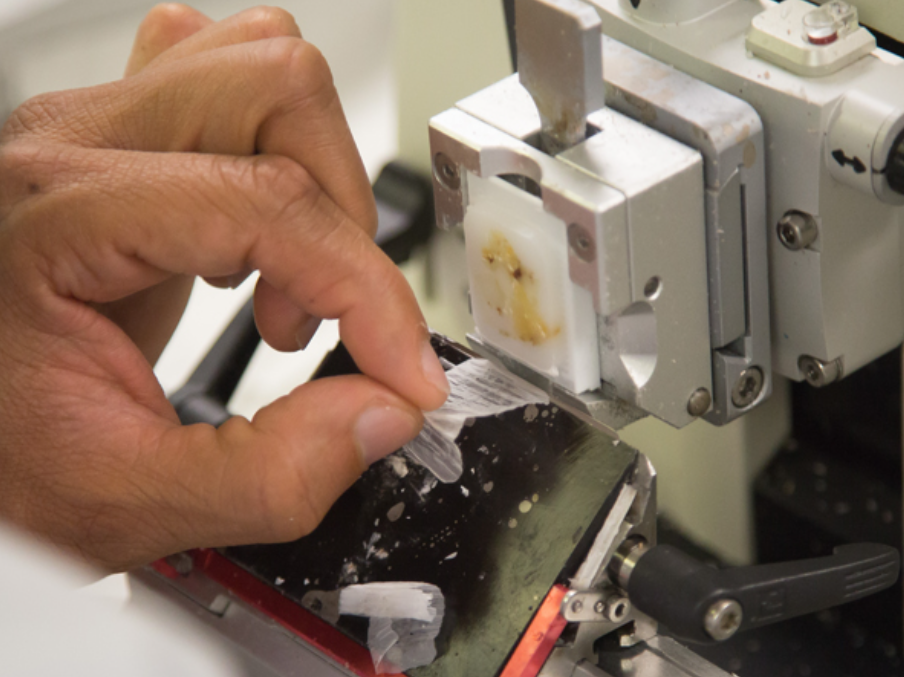
Green Light for Histotechnology Advanced Certificate
A new Advanced Certificate in Histotechnology Program will take a step toward solving the workforce shortage of state-licensed histotechnologists.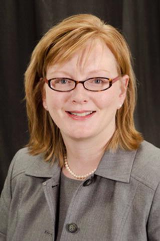
Christa Whitney-Miller Named Chair of Pathology and Laboratory Medicine
The professor of Pathology and Laboratory Medicine and vice chair of Anatomic Pathology at the University of Rochester Medical Center, has been selected as department chair and named the Frieda Robscheit-Robbins Professor after a nationwide search.
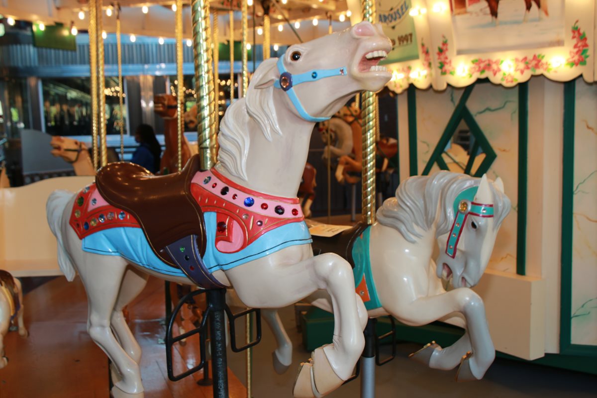
Nearly 800 Attend Employee Appreciation Event at Strong Museum of Play
Nearly 800 faculty, staff, students, and trainees celebrated the department's teamwork and success over the past year.

Welcome to our new Ph.D. students
We are happy to announce the arrival of our incoming class of Pathology Program Ph.D. students.

A Summer of Learning
Our Explorations in Pathology pre-college program each summer introduces 10 high school juniors and seniors to pathology and the many career path opportunities it presents.