
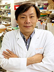
Chang Receives Patent for Prostate Cancer Treatment Method
This month, Chawnshang Chang, Ph.D. received a U.S. patent for a new way to treat prostate cancer.
His methods aim to both treat prostate cancer patients and keep it from coming back.
In his description Chang notes that patients who are treated with the commonly used method of androgen deprivation therapy (ADT) often experience a return, even after remission.
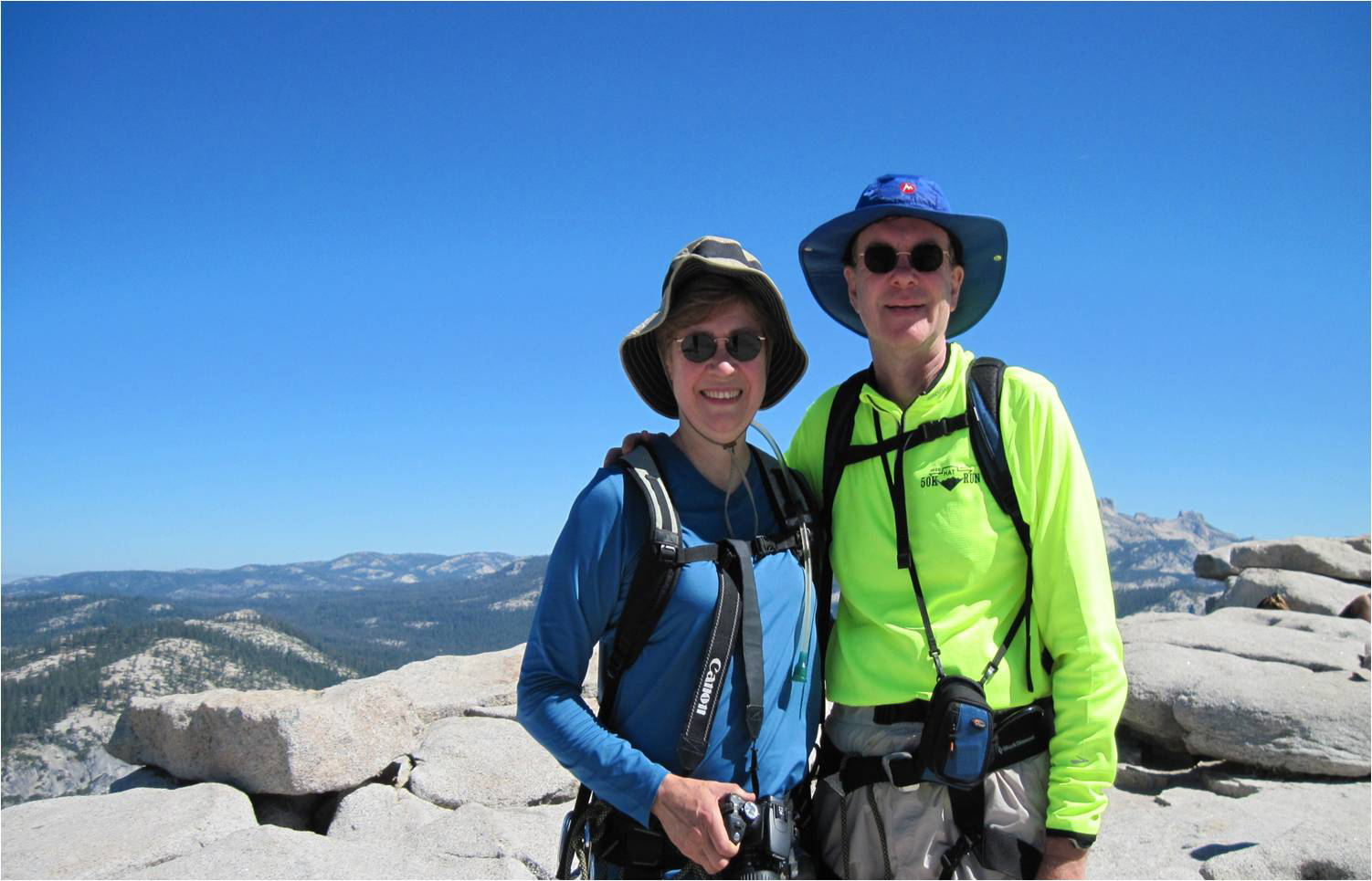
Meet UR Pathology Alumni, Drs. David Wilbur and Margaret Fallon
Drs. David Wilbur and Margaret Fallon first crossed paths during medical school at the University of Rochester, from which both graduated in 1980, after year-out student fellowships in pathology, and later joined as faculty members.
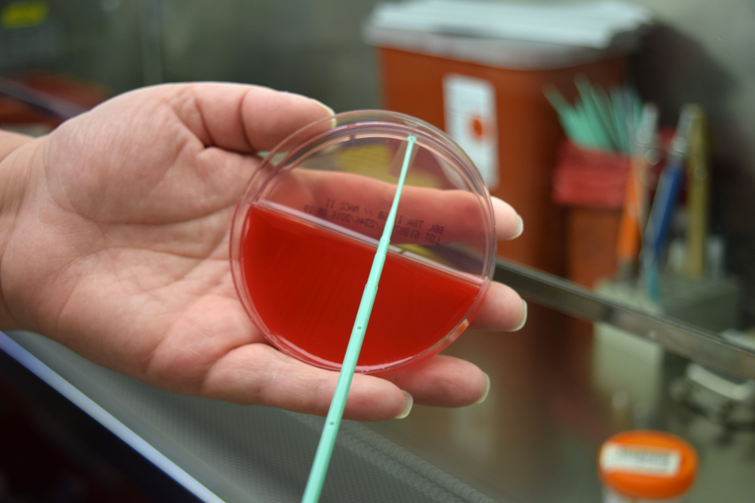
A Look Inside the Bacteriology Lab: What Does Your Culture Grow?
If you’ve ever had a sore throat swabbed to test for strep, you have experienced just one way bacteria cultures can be used to help sick patients get answers.
The Bacteriology Laboratory at Strong Memorial Hospital runs hundreds of tests around the clock to identify the bacteria and fungi that cause everything from urinary tract infections to food poisoning. This identification process is the first step in stopping sickness in its tracks and putting patients on the road to recovery.