Incidental Blasts in Peripheral Blood
Incidental Blasts in Peripheral Blood
Tausif A Siddiqui, BS; Hibbah Nabeel, MBBS
Department of Pathology, University of Rochester Medical Center, Rochester, NY, USA
Clinical History
A 77-year-old male with a history of carotid artery stenosis status post right carotid endarterectomy, benign prostatic hyperplasia, hypertension, and hyperlipidemia presented to his primary care physician after returning from a trip to Vietnam in February 2026. He reported persistent constipation, decreased appetite, fatigue, and an unintentional 10-pound weight loss over one month. He denied fever, nausea, vomiting, chest pain, dyspnea, or bleeding symptoms. Initial laboratory evaluation revealed mild anemia with circulating blasts on differential, prompting further hematologic workup.
Recent Medical History
Initial laboratory evaluation demonstrated mild macrocytic anemia with a hemoglobin of 11.1 g/dL and mean corpuscular volume of 99. The white blood cell count was 6.9 x 10³/µL and platelet count was 169 x10³/µL.
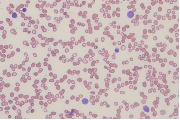
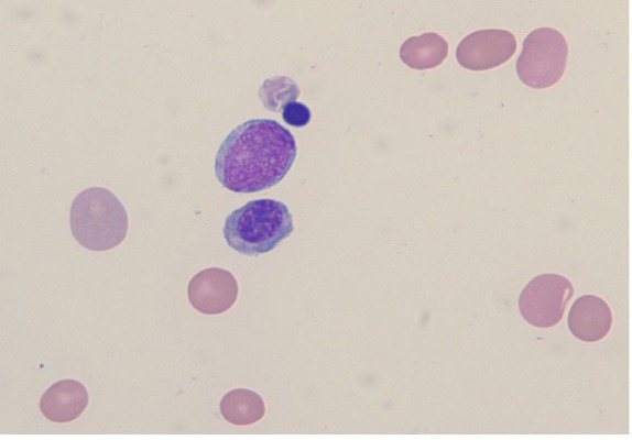
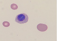
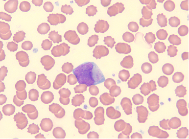
Peripheral blood smear showed anisocytosis with ovalocytes, teardrop cells, and nucleated red blood cells. Neutrophils demonstrated left-shifted maturation up to metamyelocytes with hypogranular forms and occasional hypolobation. Platelets were adequate. Increased circulating blasts were identified. These were large cells with a high nuclear-to-cytoplasmic ratio, fine chromatin, prominent nucleoli, and scant basophilic cytoplasm. Occasional blasts contained Auer rods, and some demonstrated nuclear indentations suggestive of monocytic differentiation.
Flow cytometry identified a blast population comprising around 62% of total events, expressing CD34 (partial), HLA-DR, cytoplasmic MPO (cyMPO), CD117, CD13, CD33, CD38, CD123 (dim), and CD7, with a subset expressing CD11b. Blasts were negative for TdT, CD56, CD3, CD19, CD20, and CD79a.
Fluorescence in situ hybridization (FISH) studies were negative for RUNX1T1::RUNX1 rearrangement (t(8;21)) and PML::RARA rearrangement (t(15;17)). Cytogenetic analysis demonstrated two related abnormal clones, including a stemline with a marker chromosome and a sideline clone with trisomy 4.
Next-generation sequencing using a myeloid mutation panel identified pathogenic variants in TET2 and PHF6, including a frameshift mutation in TET2 (p.Val386SerfsTer4) and a nonsense mutation in PHF6 (p.Arg129Ter), along with additional splice-site variants involving both genes. PCR testing for FLT3 internal tandem duplication and tyrosine kinase domain mutations was negative.
Following these findings, the patient was referred to hematology and subsequently underwent bone marrow aspiration and biopsy from the left posterior iliac crest for further diagnostic evaluation.