Breast MRI
Make Appointments & Get Care
What Is a Breast MRI?
A breast MRI (magnetic resonance imaging) creates detailed images of the breast tissues using large magnets and a computer. Unlike X-rays, MRI does not use radiation. Providers often use breast MRIs to check known breast cancer. Breast MRIs are often used with mammograms and ultrasounds to help find and diagnose breast cancer and other breast health issues.
How Are Breast MRIs Different from Mammograms and Ultrasounds?
Breast MRIs use magnetic fields and radio waves to create images of the breast, whereas mammograms use X-rays and ultrasounds use high-frequency sound waves to generate images.
These imaging tests are also used for different purposes:
Breast MRI
- Used for high-risk patients to assess the extent of cancer.
- Evaluate abnormalities that are not clearly visible on mammograms or ultrasounds.
- Not recommended as a stand-alone screening test, as it can miss cancers that a mammogram would find.
Mammograms
- Used for routine breast cancer screening
- Can detect tumors that are too small to be felt.
Ultrasounds
- Used to further evaluate abnormalities found on a mammogram or physical exam.
- Can distinguish between solid masses and fluid-filled cysts.
Why is a Breast MRI Performed?
Breast MRIs are most commonly used to detect early breast cancers not detected by other tests, especially in high-risk patients. Your provider may order this advanced imaging to examine, evaluate, or monitor the following:
- Areas of concern seen on a mammogram
- Dense breast tissue and scar tissue
- Silicone gel breast implant leakage
- The size and location of lesions or lumps
- Lumpectomy sites after treatment
- Effectiveness of chemotherapy
- Cause of a newly inverted nipple
UR Medicine's Approach
Breast MRIs are especially important because early detection is key when it comes to diagnosing and treating breast cancer. UR Medicine’s Comprehensive Breast Center is home to some of the nation’s top breast experts. Our providers specialize in detecting, treating, and supporting patients with breast cancer. We will walk you through every step from diagnosis to treatment.
When Should I Get a Breast MRI?
The American Cancer Society recommends breast MRI and a mammogram starting at about age 30 for individuals at high risk for breast cancer, including:
- Those with a BRCA1 or BRCA2 gene mutation
- Individuals with a first-degree relative with a BRCA1 or BRCA2 gene mutation
- Those with a 20% to 25% or greater lifetime risk for breast cancer based on family history and other factors
- Individuals who had radiation treatment to the chest between ages 10 and 30
- Those with genetic disorders such as Li-Fraumeni syndrome, Cowden syndrome, or Bannayan-Riley-Ruvalcaba syndrome
- Individuals with a close family member who has one of the above genetic disorders
Some individuals should not have an MRI, including:
- Pregnant individuals
- Those with pacemakers and other implanted devices
- Individuals with recently placed metal plates, rods, screws, or other surgical devices
What Happens During a Breast MRI?
The MRI scanner is a large machine with a tunnel, and you will lie on a table that slides in and out of this tunnel. For a breast MRI, you will lie face down with your breasts positioned through holes in the table. Often, a contrast dye is injected into a vein in your arm before or during the procedure to help create clearer images.
A breast MRI can be done as an outpatient or as part of a longer hospital stay. The process typically includes:
- Preparation: You will remove your clothes and wear a hospital gown or scrubs. Remove jewelry or other objects.
- IV Line: If contrast dye is used, an IV line will be started in your hand or arm.
- Positioning: You will lie face down on a mobile bed with your breasts positioned through cushioned openings. The bed will move into the MRI machine.
- Communication: The technologist will be in a separate room but will maintain constant communication with you through speakers.
- Noise Reduction: You will wear earplugs or a headset to block out the noise from the scanner.
- Imaging: You will need to lie still and may be asked to hold your breath at times. If contrast dye is used, you may feel a flushing sensation, coldness, a metallic taste, a brief headache, itching, nausea, or vomiting.
- Completion: After the scan, the table will slide out, and you will be helped off the table. The IV will be removed if used.
What Sets Us Apart?
The UR Medicine Breast Imaging Center is designated a Breast Imaging Center of Excellence by the American College of Radiology's (ACR) Commission on Quality and Safety and the Commission on Breast Imaging for a three-year term. The ACR Breast Imaging Center of Excellence designation signifies that UR Medicine Breast Imaging provides these services to the Rochester and surrounding communities at the highest standards of the radiology profession.
Our expertise is backed by research. As part of an academic medical center, our providers are continuously working to advance the detection and assessment of breast disease. We invest in cutting-edge technology.
We collaborate with breast specialists at UR Medicine's Wilmot Cancer Institute to ensure that all necessary treatments and support is available.
Locations
View All LocationsWe serve you in the Rochester metropolitan area and surrounding region.
View All Locations6 locations
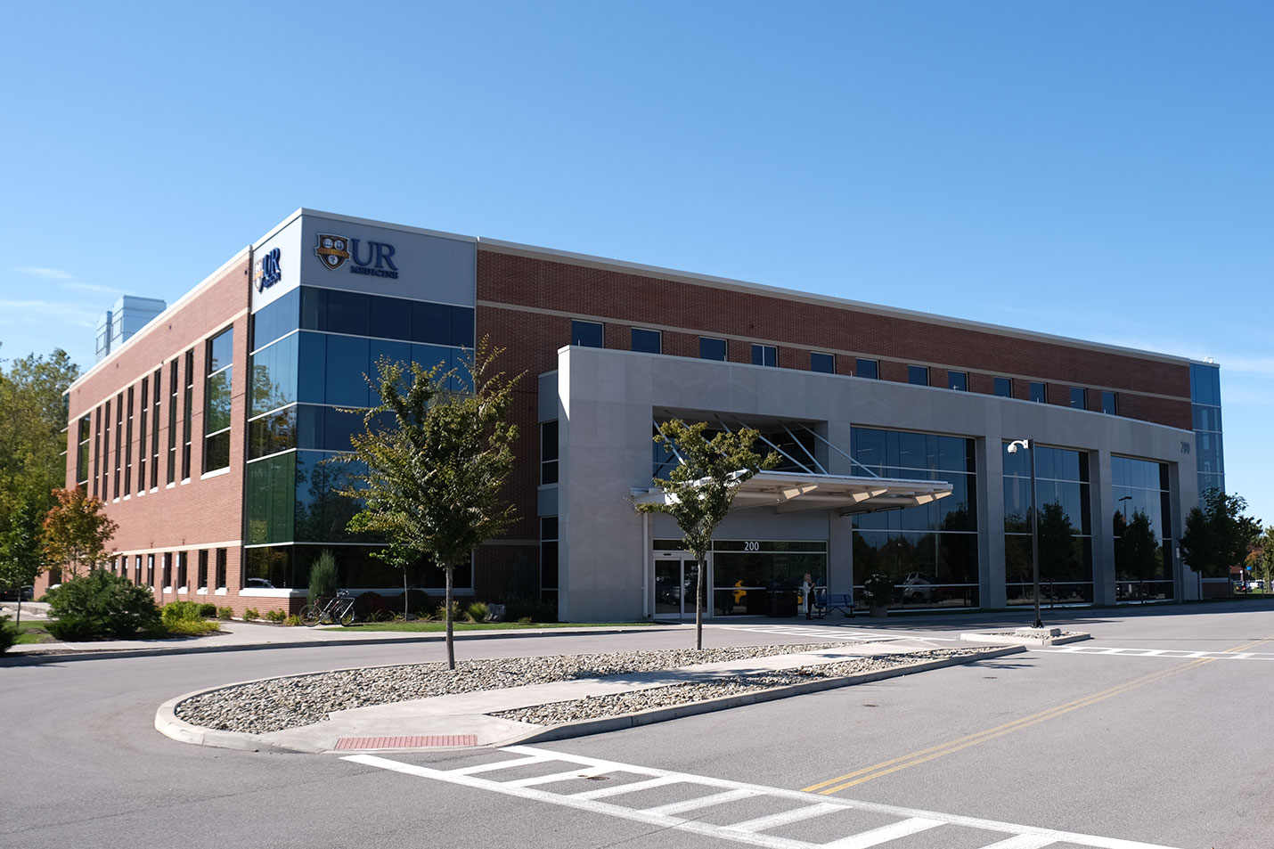
200 East River Road
Rochester, NY 14623
St. James Medical Office Building
7309 Seneca Road North, Entrance E, Suite 113
Hornell, NY 14843
Lakeside Professional Park
229B Parrish Street, Suite 103
Canandaigua, NY 14424
Strong Memorial Hospital
601 Elmwood Avenue
Rochester, NY 14642
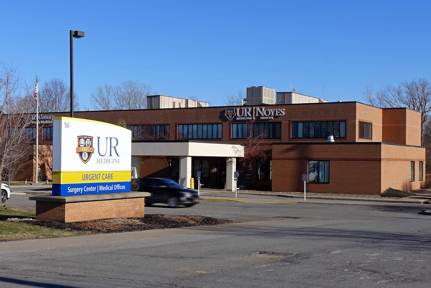
Noyes Health Services
50 East South Street
Geneseo, NY 14454
St. James Medical Office Building
7309 Seneca Road North, Entrance E, Suite 113
Hornell, NY 14843


