Breast Ultrasound
Make Appointments & Get Care
What Is a Breast Ultrasound?
A breast ultrasound is a non-invasive imaging test that uses high-frequency sound waves to create detailed images of the structures within the breast. It is used to help check for breast abnormalities, especially in telling the difference between solid masses and fluid-filled cysts.
UR Medicine offers two types of breast ultrasounds:
- Handheld Breast Ultrasound A technician uses a small handheld device, called a transducer, to move gently over your breast and capture internal images.
Breast Ultrasounds vs. Mammograms
A mammogram uses X-rays to take a clear picture of the breast, while a breast ultrasound uses sound waves. This is especially helpful for women with dense breast tissue. Usually, providers recommend a mammogram as the main screening tool, and an ultrasound is used as an extra test in certain cases. A Breast MRI may also be recommended to evaluate abnormalities that aren’t clearly visible on mammograms or ultrasounds.
Ultrasounds
- Use sound waves to create images
- Often performed on a small area due to symptoms or to evaluate findings on a mammogram (“targeted ultrasound”)
- Examine dense breast tissue (“screening ultrasound”) for cancer that may be missed by mammograms
Mammograms
- Use X-rays to create images
- Standard screening test for breast cancer
- Detect calcification and tumors
Yes, breast ultrasounds can be more effective than mammograms for women with dense breast tissue. Dense breasts have more fibrous and glandular tissue, which can make it difficult for mammograms to detect abnormalities. Ultrasounds provide a clearer image of dense tissue, helping to identify potential issues that mammograms might miss.
Yes, breast ultrasounds can help detect cancer, especially in dense breast tissue where mammograms might be less effective. Ultrasounds are not typically used as a primary screening tool for breast cancer, but they are valuable for further evaluating mammogram findings and for guiding biopsies. However, they may not detect all types of breast cancer, so they are often used alongside mammography or breast MRIs.
Breast ultrasound is often used to:
- Investigate a detectable lump in the breast
- Screen for breast cancer in dense breast tissue
- Evaluate abnormalities found on a mammogram
- Guide needle biopsies
- Monitor changes in breast tissue over time
- Assess breast implants
UR Medicine's Approach
Early detection is key in diagnosing and treating breast cancer, and breast ultrasounds play an important role in early detection. UR Medicine’s Comprehensive Breast Center is home to some of the nation’s top breast experts. Our providers specialize in detecting, treating, and supporting patients with breast cancer. We will walk you through every step from diagnosis to treatment.
A breast ultrasound typically takes 15 to 30 minutes. During the procedure, you will lie on your back or side on an examination table. You may be asked to change positions to get different views. A technician will apply a water-based gel to your skin and move the transducer over the breast to capture images.
Preparation for a breast ultrasound is minimal. Here are some general guidelines:
- Wear comfortable clothing that can be easily removed or adjusted.
- Avoid applying lotions, powders, or deodorants on the day of the exam, as these can interfere with the ultrasound images.
- Inform your healthcare provider of any recent breast surgeries or procedures.
A radiologist will analyze the ultrasound images and provide a report to your healthcare provider. Your provider will discuss the results with you and explain any further steps if necessary.
Possible findings include:
- Cysts: Fluid-filled sacs that are usually benign.
- Solid Masses: These may require further evaluation to determine if they are benign or malignant.
- Other Abnormalities: Any unusual findings will be assessed for further diagnostic testing.
What Sets Us Apart?
The UR Medicine Breast Imaging Center is designated a Breast Imaging Center of Excellence by the American College of Radiology's (ACR) Commission on Quality and Safety and the Commission on Breast Imaging for a three-year term. The ACR Breast Imaging Center of Excellence designation signifies that UR Medicine Breast Imaging provides these services to the Rochester and surrounding communities at the highest standards of the radiology profession.
Our expertise is backed by research. As part of an academic medical center, our providers are continuously working to advance the detection and assessment of breast disease. We invest in cutting-edge technology.
We collaborate with breast specialists at UR Medicine's Wilmot Cancer Institute to ensure that all necessary treatments and support is available.
Locations
View All LocationsWe serve you in the Rochester metropolitan area and surrounding region.
View All Locations10 locations
St. James Medical Office Building
7309 Seneca Road North, Entrance E, Suite 113
Hornell, NY 14843
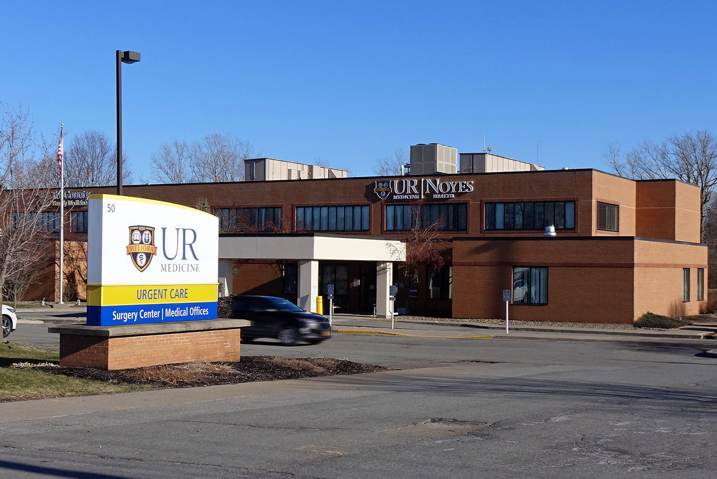
Noyes Health Services
50 East South Street
Geneseo, NY 14454
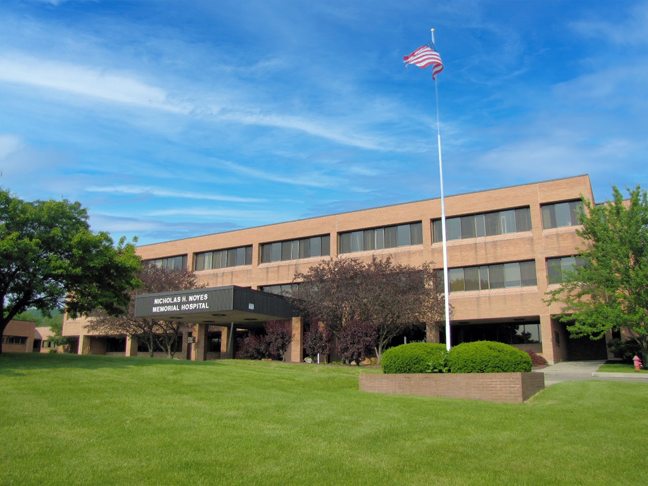
E. Michael Saunders Medical Imaging at Noyes Memorial Hospital
111 Clara Barton Street
Dansville, NY 14437
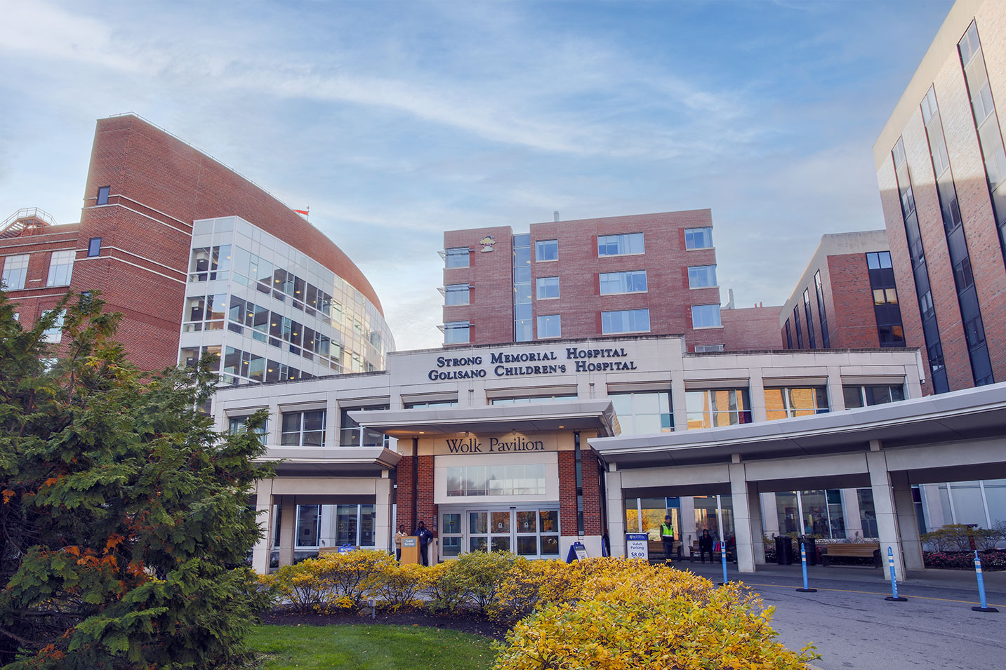
Strong Memorial Hospital
601 Elmwood Avenue
Rochester, NY 14642
Calkins Corporate Park (Red Creek)
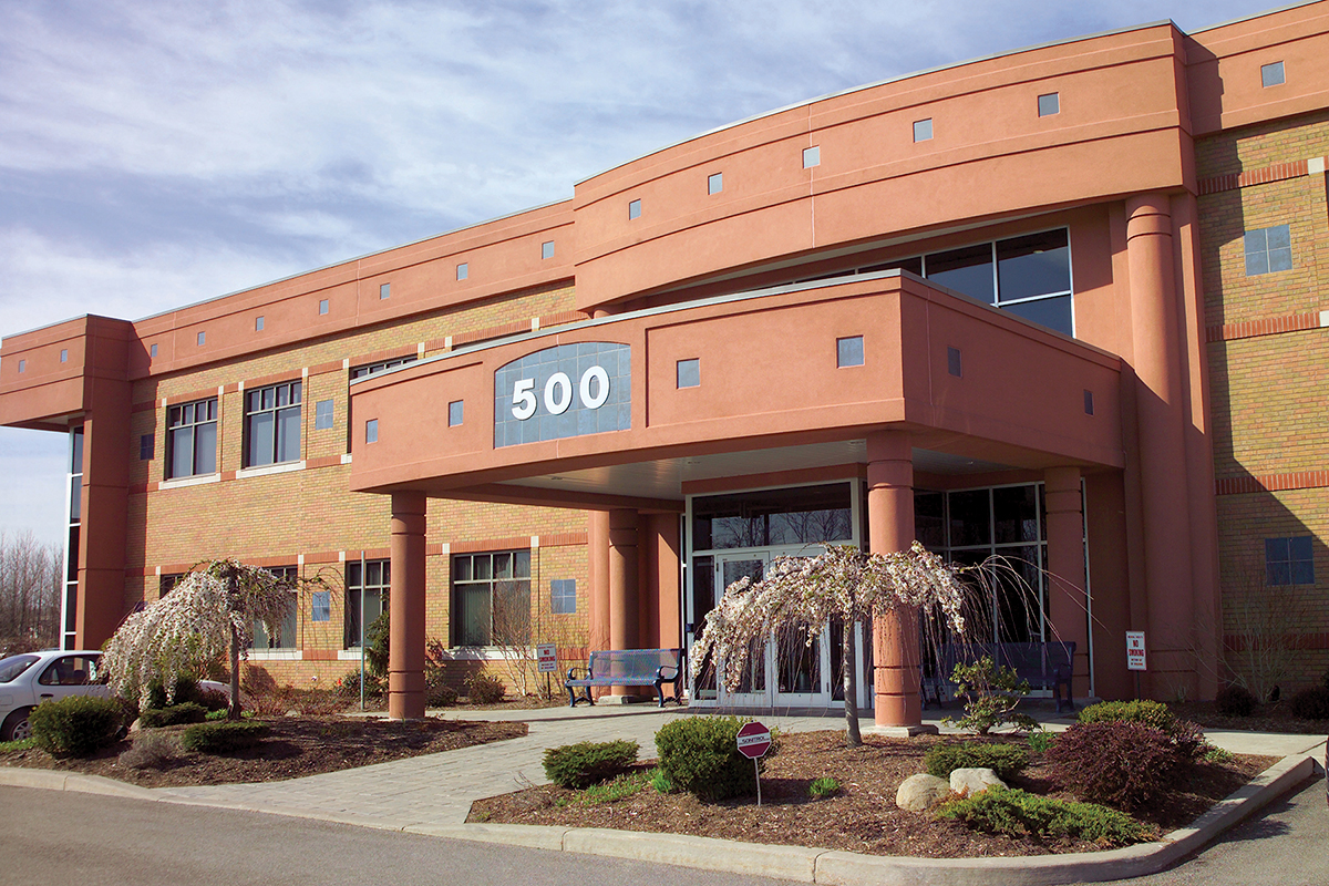
500 Red Creek Drive, Suite 130
Rochester, NY 14623
Lakeside Professional Park
229B Parrish Street, Suite 103
Canandaigua, NY 14424
Strong Memorial Hospital
601 Elmwood Avenue
Rochester, NY 14642
E. Michael Saunder Medical Imaging at Noyes Memorial Hospital
111 Cara Barton Street
Dansville, NY 14437
Noyes Health Services
50 East South Street
Geneseo, NY 14454
St. James Medical Office Building
7309 Seneca Road North, Entrance E, Suite 113
Hornell, NY 14843
