

News from the Dalecki Lab
News from the Dalecki Lab
2024 RCBU Biomedical Ultrasound Symposium Day
Tuesday, November 12, 2024
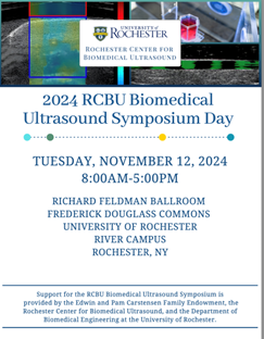 The 2024 RCBU Biomedical Ultrasound Symposium was an exciting day dedicated to biomedical ultrasound research and technology! Each year, the RCBU Symposium brings together faculty, researchers, trainees, colleagues from other universities, and industry partners to share the latest research advances in ultrasound. The symposium consists of Distinguished Lectures, scientific presentations, poster presentations, and networking all dedicated to biomedical ultrasound. This year’s symposium featured four Keynote lectures:
Keynote Lectures:
Distinguished Edwin L. Carstensen Lecture
“Quantitative Imaging Biomarkers: Medical Imaging Meets Metrology”
Timothy J. Hall, PhD, University of Wisconsin-Madison
Distinguished RCBU Alumni Lecture
“Enabling Ultrasound Techniques for Tissue Characterization”
Karla P. Mercado-Shekhar, PhD, Indian Institute of Technology (IIT) Gandhinagar
Clinical Challenges
“Histotripsy for Liver Malignancy”
Koji Tomiyama, MD, University of Rochester
Hot Topic Keynote
“A Focused Ultrasound Therapy for More than Just Tissue Ablation”
Kenneth B. Bader, PhD, University of Chicago
Karla Mercado-Shekhar, PhD returns to deliver the Distinguished RCBU Alumni Lecture!
Tuesday, November 12, 2024
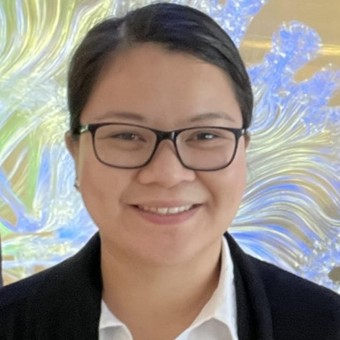 Dr. Karla Mercado-Shekhar returned to the University of Rochester to deliver the Distinguished RCBU Alumni Lecture at the 2024 RCBU Biomedical Ultrasound Symposium. Dr. Mercado-Shekhar is an alumna from the Dalecki Lab and earned her Ph.D. in Biomedical Engineering in 2015. At the RCBU Symposium, she delivered an exciting talk on her latest research titled "Enabling ultrasound techniques for tissue characterization". Professor Dalecki stated, "It was so wonderful to honor Karla’s achievements and professional success with this Distinguished RCBU Alumni Lecture!"
Karla is an Assistant Professor of Biological Sciences and Engineering at the Indian Institute of Technology (IIT) Gandhinagar. She received her B.S. in Biomedical Engineering from Boston University (2009) and her M.S. (2010) and Ph.D. (2015) in Biomedical Engineering from the University of Rochester, as a Provost’s Fellow. She was a Postdoctoral Fellow at the University of Cincinnati College of Medicine (2015-2019) and a recipient of a postdoctoral research supplement grant from the National Institutes of Health. Karla co-leads the Medical Ultrasound Engineering (MUSE) Lab where her research focuses on ultrasound tissue characterization, viscoelasticity imaging, and drug delivery. She has been recognized as a Scholar by the IEEE Ultrasonics, Ferroelectrics, and Frequency Control Society. She serves on the Advisory Editorial Board of the journal Ultrasound in Medicine and Biology, and the Technical Program Committees of the IEEE International Ultrasonics Symposium and the Biomedical Acoustics Technical Committee of the Acoustical Society of America.
Scientists leverage ultrasound to build new blood vessels in living tissue
Tuesday, September 10, 2024
The novel technique could be used to treat damaged tissue in a range of medical applications, including reconstructive and plastic surgeries
A technology most often used for medical imaging is being repurposed as a new tool for restoring blood flow in tissue damaged from disease, injury, and reconstructive surgery. Biomedical engineers at the University of Rochester are leveraging ultrasound waves to organize endothelial cells—the building blocks of blood vessels—into patterns that can promote the growth of new vessel networks within days.
“We developed a novel technique using some of the forces associated with an ultrasound field called acoustic radiation force to act on cells in a material to move them to different spatial locations,” says Diane Dalecki, the Kevin J. Parker Distinguished Professor in Biomedical Engineering and director of the Rochester Center for Biomedical Ultrasound. “By changing the frequency of the sound fields, we can control the distance between how the cells are patterned. Depending on the patterning we use, we can create different types of blood vessel morphologies.”
A team led by Dalecki and Denise Hocking, a professor of pharmacology and physiology and of biomedical engineering, have used the technique to engineer tissue with new blood vessel networks in vitro. In their recently published studies, they showed that acoustic patterning can also be used to produce new blood vessels directly in the body. A new $2 million grant from the National Institutes of Health will help the researchers refine their in vivo acoustic patterning technologies.
“Rather than making an engineered tissue product outside of the body and then implanting it, we would like to induce the formation of new blood vessels directly in the body,” says Hocking. “Ultrasound has the ability to penetrate through tissue and is already used in many clinical applications, so why not try to produce new vessels locally?”
Read More: Scientists leverage ultrasound to build new blood vessels in living tissueScientists Leverage Ultrasound to Build New Blood Vessels in Living Tissue
Tuesday, September 10, 2024
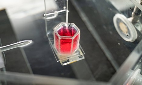 A technology most often used for medical imaging is being repurposed as a new tool for restoring blood flow in tissue damaged from disease, injury, and reconstructive surgery. Biomedical engineers at the University of Rochester are leveraging ultrasound waves to organize endothelial cells—the building blocks of blood vessels—into patterns that can promote the growth of new vessel networks within days.
Professors Dalecki and Hocking Receive New NIH Grant
Tuesday, July 16, 2024
Professor Diane Dalecki (BME) and Professor Denise Hocking (Pharmacology & Physiology) received a new, NIH R01 grant titled “In Vivo Acoustic Patterning for Tissue Vascularization”. This Multi-PI award will enable the team to develop novel ultrasound technologies for tissue engineering and regeneration. The overall goal of the project is to advance acoustic patterning as a noninvasive, versatile approach to produce functional microvascular networks directly in vivo. The advantage of this technology is the ability to focus ultrasound fields within the body noninvasively and generate three-dimensional patterns of cells suspended within injectable hydrogels site specifically. The team’s ultrasound-based patterning technology relies on the use of acoustic radiation forces generated within standing wave fields, or acoustic fields from holographic lens transducers, to rapidly and noninvasively pattern cells within collagen-based hydrogels. Dr. Mohamed Ghanem (University of Washington, Seattle) also joins the team and brings his expertise on design and fabrication of holographic lens transducers and modeling acoustic fields and radiation forces generated by these unique acoustic sources. The grant is sponsored by the National Institute of Biomedical Imaging and Biomedical Engineering (NIBIB) within the National Institutes of Health (NIH).