Research Projects
Clinical Projects
The Use of Diffusion Magnetic Resonance Imaging to Characterize the Intra-cranial Path of the Facial Nerve
The goal of this study was to develop a high-resolution MRI and diffusion MRI (dMRI) protocol to identify the intra-cranial course of the facial nerve.
While the majority of Bell’s Palsy (BP) patients recover full function of the facial nerve (FN), a small percentage remain fully paralyzed. Traditionally, diagnosis is one of exclusion, and those that fail to recover are often observed for extended periods in the hope of some functional recovery. This approach often renders the existing facial musculature unusable due to irreversible end-plate degeneration. Currently there is no investigation that identifies the BP patients won’t fully recover. These patients may benefit from earlier surgical intervention to utilize the existing facial muscles.
We have developed a novel technique to automatically quantify facial asymmetry in videos for patient treatment and rehabilitation. A state-of-the-art landmark detection technique is effective for style invariant landmark detection. Our preliminary data suggests this method may provide a subjective and accurate analysis of facial nerve recovery.
Developing an Automated Facial Symmetry Index (AFSI)
The goal of this project is to evaluate the automated facial symmetry index (AFSI) tool to assess facial symmetry that can be used to monitor the recovery of facial movements.
The ideal functional outcome measurement for facial nerve injury continues to remain controversial. Ideally, a subjective, minimally invasive, and easily implemented tool would be available. While some automated systems have already been developed and validated, they are largely 2-D renderings, subjective in nature, and capture only static images thus failing to incorporate the dynamism of facial movements.
We developed a novel technique to automatically quantify facial asymmetry in videos for patient treatment and rehabilitation. A state-of-the-art landmark detection technique is effective for style invariant landmark detection. Preliminary data suggests this method may provide an objective and accurate analysis of facial nerve recovery.
QoL and Surgical Outcomes of Vascularized Composite Allotransplantation
24 years after the first successful upper extremity transplantation, the role of vascularized composite allotransplantation (VCA) in the world of transplantation remains controversial. Face and upper extremity reconstruction via transplantation have become successful options for highly selected patients with severe tissue and functional deficit when conventional reconstructive options are no longer available. Despite clear benefit in these situations, VCA has a significant potential for complications that are more frequent when compared to visceral organ transplantation.
Our focus is to look at novel ways to manipulate the immune system to reduce the severity of rejection.
Basic Science
Prevention and Treatment of Chemotherapy Induced Peripheral Neuropathy
The over-arching hypothesis of this project is that the anti-inflammatory response of type 2 macrophages and pro-myelinating response of increased c-jun expression will mitigate the development of CIPN.
An estimated 30-70% of the 650,000 of patients treated with chemotherapy every year in the United States will develop symptoms of CIPN that range from numbness and tingling in the hands and feet, to burning pain, muscle weakness and loss of co-ordination. CIPN is a leading cause for therapeutic non-compliance, reduced quality of life and poorer cancer survival rates. Patients who develop symptomatic CIPN also have an associated $20,000 increase in overall treatment cost in comparison to patients that do not develop symptoms and are also more likely to suffer relapse of their cancer.
One of the cancers in which CIPN is well-studied is breast cancer, which was the most common cancer in women worldwide in 2018, accounting for more than a quarter of all cancers. Surgery to remove tumors and affected lymph nodes is effective in early disease, but the mainstay of treatment in resistant tumors is with neoadjuvant chemotherapy. The preferred chemotherapy agents used in this patient group are taxanes of which Paclitaxel (PTX) is most common. PTX is an effective agent at treating hormone resistant metastatic breast cancer. The problem is that PTX frequently induces CIPN. Currently, there are no established treatments available for CIPN patients. Pre-clinical studies are investigating multiple compounds to prevent or treat CIPN by blocking ion channels, targeting inflammatory cytokines, and combating oxidative stress, yet the results have not been encouraging.
Murphy Roths Large (MRL/MpJ) is the parent and control strain for MRL/MpJ-Faslpr. MRL/MpJ-Faslpr carry mutation in Fas gene and therefore both strains were originally developed as a model for autoimmune disorders. MRL/MpJ has demonstrated superior healing features with minimal or even scar-less wound healing in certain body areas. First report of enhanced healing in MRL/MpJ occurred after a spontaneous closure of ear holes used for mouse identification. Priorly, we have shown that these beneficial healing effects also confer to the regenerating peripheral nervous system. MRL/MpJ mice demonstrate superior functional outcomes that, in part, can be attributed to improved extracellular matrix (ECM) remodeling, that is driven by an increased activity of M2a macrophages and neutrophils. Additionally, the MRL/MpJ strain demonstrate increased myelination of regenerating axons that is likely driven by an upregulation of c-jun expression, resulting in Schwann Cell (SC) preferentially trans-differentiating into reparative SCs. Taking these into consideration, the MRL/MpJ strain may provide: 1) An anti-inflammatory effect that reduces the impact of ROS through increased activity of type 2a macrophages and 2) Through the upregulation of c-jun, protection against demyelination of peripheral axons. Taken in combination, these independent processes may provide targets that mitigate the onset of CIPN.
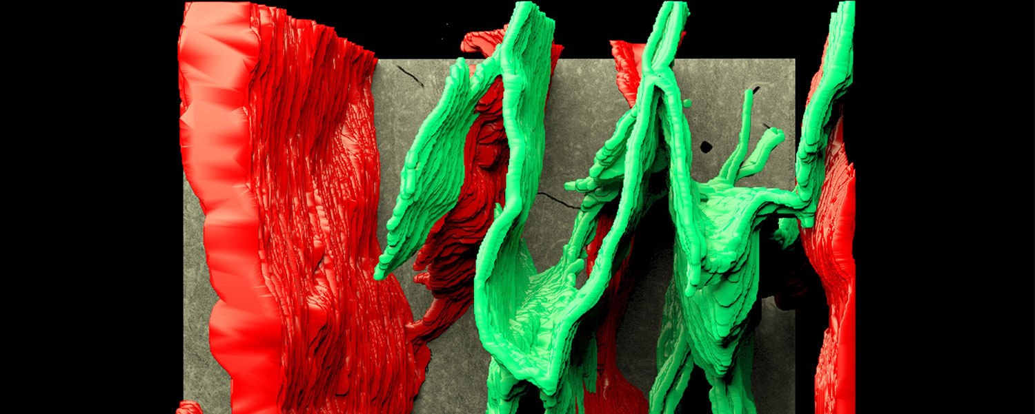
Application of Serial Section Microscopy (SSEM) to Evaluate Small Tissue Volumes in Disease
SSEM involves a continuous collection of ultra-thin sections onto a reel of Kapton® tape using an automated tape-collecting ultramicrotome (ATUMtome, RMC Boeckeler, USA). Each section is imaged in sequential order using a backscatter electron detector (BSE), to produce a ‘stack’ of images that can be analyzed to examine the cellular processes within the volume of tissue processed.
The lab has completed exploratory investigations to apply this process to the regenerating peripheral nervous system to increase our understanding of axonal regeneration. Similarly, collaborative efforts with the Center for Musculoskeletal Research, we have been able to visualize the colonization of the canalicular network in bone infected with S. aureus in three dimensions.