Photo Contest Selections
Photo Contest Selections
General Information
Send us your most creative scientific works of art and be entered into a "peoples choice" award for best image.
Please submit a single file in PDF format which includes a brief (400 character max) description of your work and a photo (4"x6" high resolution) that you would like for the contest.
Only a single entry will be accepted per individual.
Submitted Photos
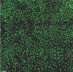
1:
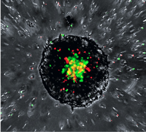
This image shows neutrophils actively swarming and infiltrating a pancreatic cancer tumoroid. Green cells are neutrophils derived from tumor bearing mice while red cells are neutrophils derived from naïve mice. Yellow depicts the proximal overlap between the two neutrophil types
This image shows neutrophils actively swarming and infiltrating a pancreatic cancer tumoroid. Green cells are neutrophils derived from tumor bearing mice while red cells are neutrophils derived from naïve mice. Yellow depicts the proximal overlap between the two neutrophil types
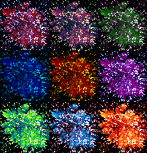
2:
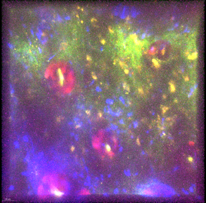
Psychedelic Immunity
Psychedelic Immunity
3:
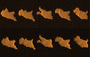
3D visualization of a T cell hybridoma migrating between two polyacrylamide gel. The top row is the top view and the bottom row is the bottom view of the cell. Going from left to right is five different time point, each of which are separated by 3.5 min. The bump observed under the cell and slowing appearing on the top of the cell are focal adhesions that are forming as the cell migrates
3D visualization of a T cell hybridoma migrating between two polyacrylamide gel. The top row is the top view and the bottom row is the bottom view of the cell. Going from left to right is five different time point, each of which are separated by 3.5 min. The bump observed under the cell and slowing appearing on the top of the cell are focal adhesions that are forming as the cell migrates
4:
MHC Class2 Expressing Langerhans Cells
H2-Eb1-Ametrine Expressing Mouse
MHC Class2 Expressing Langerhans Cells
H2-Eb1-Ametrine Expressing Mouse
5:
Periodic acid Schiff stain (with diastase pretreatment) of a rhabdoid tumor
Periodic acid Schiff stain (with diastase pretreatment) of a rhabdoid tumor
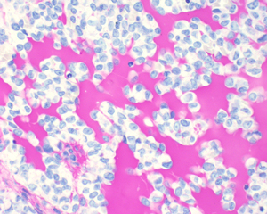
6:
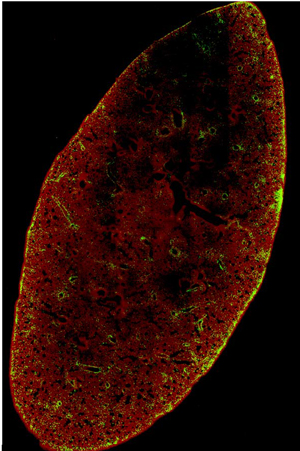
Deep 3D imaging (1 mm thick) of CAR-T cell accumulation in a cleared mouse lung 72 h after i.v. injection (red; blood vessels [CD31], green; CAR-T cells [GFP])
Deep 3D imaging (1 mm thick) of CAR-T cell accumulation in a cleared mouse lung 72 h after i.v. injection (red; blood vessels [CD31], green; CAR-T cells [GFP])
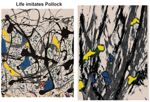
7:
8:
9: