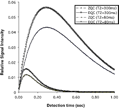
It was found that in general the intensity of iMQC of different orders
signals are only a few percent of the intensity of the SQC, but iDQCs
have a sensitivity about 30% higher than iZQC for human imaging.
This was confirmed with experiments in uniform phantoms and in human
brain tissue at 1.5T. The duration of iMQC signals is found to be much
greater than that of SQCs with the same T2. Also the maximum of the
signal is centered at a later time.
We are among the first to develop intermolecular multi-quantum coherence (iMQC) imaging techniques, showing theoretical properties of related signals, illustrating its potential applications in variety of imaging and spectroscopic applications, and suggesting its potential for fMRI with more specific characteristics at high field.
Our research in the area has resulted in over 20 publications, and issue of an US patent (#6,528,997). In the past few years we have collaborated with other researchers to develop spatial encoding (SPEN) techniques for novel imaging applications, such as fast imaging of water and fat in brain.
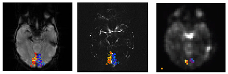
Results from experiments with presentations of hemifield visual stimulus that
last 30 sec for the left side, then 30 sec for the right side, and repeat 24 times
for iDQC, and 8 times for the conventional SQC BOLD. TR is 5 sec for iDQCand
3s for SQC. (Left): GE- SQC activation pattern superimposed on the raw EPI
images in a single subject. Hot and cool colors represent activations to separate
hemifield stimulations. (Middle) The same activation pattern (interpolated to higher
resolution) superimposed on the MR angiograph show locations of major blood
vessels. (Right): SE-iDQC activation map of the same slice. Note the less
susceptibility related distortion in iDQC image due to its spin-echo nature
in readout. Robust activation is however detected. These observations are
consistent among 5 subjects studied so far. Locations of activations from both
types of images share some common voxels (note the cross hair on both) but
fewer voxels are activated in iDQC, which may reflect its lower SNR and/or
spatial resolution, but could also be due to selection of iDQC to different
vessel scales.