Equipment
Equipment
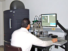
 The IVIS® Spectrum has unique capabilities for sensitively imaging both bioluminescent and fluorescent reporters within the same animal without mixing the multi-spectra. The system performs both epi- and trans-illumination fluorescent imaging and uses high efficiency narrow band-pass filters coupled with spectral unmixing algorithms to differentiate between multiple shallow and deep fluorescent sources. This is useful in optimally enabling a wide array of fluorescence/bioluminescence research applications. The system is also capable of performing 3D tomographic reconstructions for both bioluminescence and fluorescence, and enables data co-registration with complementary modalities such as CT and MRI.
Specific Features in the IVIS® Spectrum Instrument
- Transmission Fluorescence Imaging
- Excitation light originating underneath the stage trans-illuminates the subject at precise x,y-locations, enabling transmission imaging. Transmission fluorescence imaging enables more sensitive detection, accurate quantification of deep sources, and reduced auto-fluorescence effects.
- Narrow Band Excitation and Emission Filters
- The IVIS® Spectrum excitation and emission filters enable spectral scanning over a wide spectrum from the blue to NIR wavelength region. These include:
- 10 narrow band excitation filters: 415 nm -760 nm (30 nm bandwidth)
- 18 narrow band emission filters: 490 nm -850 nm (20 nm bandwidth)
- Spectral Unmixing Algorithms
- The Living Image 3.0 software provides advanced spectral unmixing algorithms that allow detection and separation of multiple reporters, greatly reduce the effects of tissue autofluorescence, and effectively reduce cross talk between reporters.
- These narrow band excitation and emission filters and spectral unmixing algorithms allow full advantage of reporters across the blue to NIR wavelength region, with ability to visualize multiple reporters in the same subject with reduced cross talk, and with significantly reduced auto-fluorescence.
- 3D Fluorescence Imaging Tomography (FLIT)
- 3D fluorescence tomography is a method that takes into account the scattering and absorption of light in tissue, and provides an estimate of the 3D location and brightness of the light emitting source.
- The IVIS® Spectrum is equipped with a single-view 3D tomography capability, meaning the 3D reconstruction can be performed from images acquired from a single-view vantage point. Using the Spectrum’s unique trans-illumination capability, images are acquired at several different trans-illumination points to determine the localization of the source position.
- 3D Diffuse Luminescence Tomography (DLIT)
- Bioluminescence imaging, like fluorescence imaging, uses light scattering and absorption to determine depth and intensity of signal. However images are acquired at several different wavelengths to improve localization of the source position. Six 20 nm wide filters centered at 560, 580, 600, 620, 640, and 660 nm are included for this application.
- Co-Registration with Other 3D Imaging Modalities
- The Spectrum can import and automatically co-register CT or MRI images yielding a functional and anatomical context for the bioluminescence and fluorescence data.
- XGI-8 Gas Anesthesia Module
- A built-in isoflurane vaporizer with independent flow control for proper gas delivery to induction chamber and the IVIS® Imaging System imaging chamber, which includes isoflurane-absorbing disposable charcoal filters to absorb excess gas, as well as limit gas escaping into the surrounding laboratory environment, an induction chamber - anesthetizes up to 5 adult mice or 2 rats simultaneously - Ventilates excess gas automatically when lid is opened - Raised animal floor maintains animal cleanliness during anesthetization, and a 5-Port anesthesia manifold - Delivers gas to up to 5 adult mice or 2 rats.