Case of the Month: Inguinal Mass
Phoenix D. Bell, MD, Gregory A. Gates, DO, and Aaron R. Huber, DO
Clinical History
A 29-year-old healthy man presented to the outpatient surgery clinic with a left inguinal mass associated with left testicular pain. He denied any other symptoms.
Past Medical History
The patient had no significant past medical history and no previous surgeries.
Recent History
The patient underwent an excision of the inguinal lesion without complication. He has since been followed with yearly imaging and has had no recurrence of the mass.
Pathology
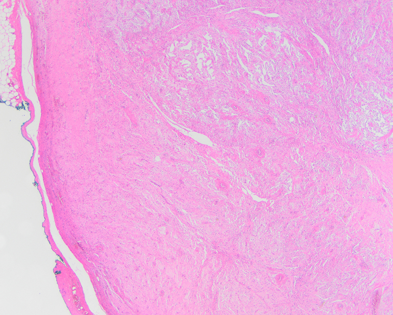
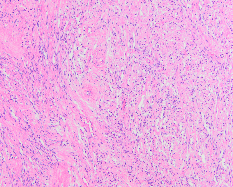
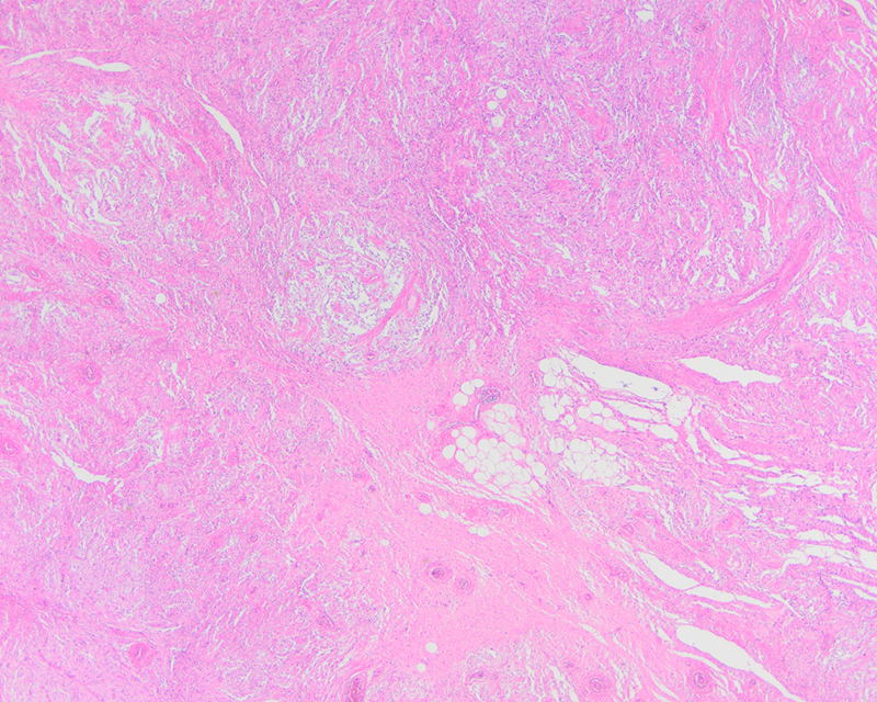
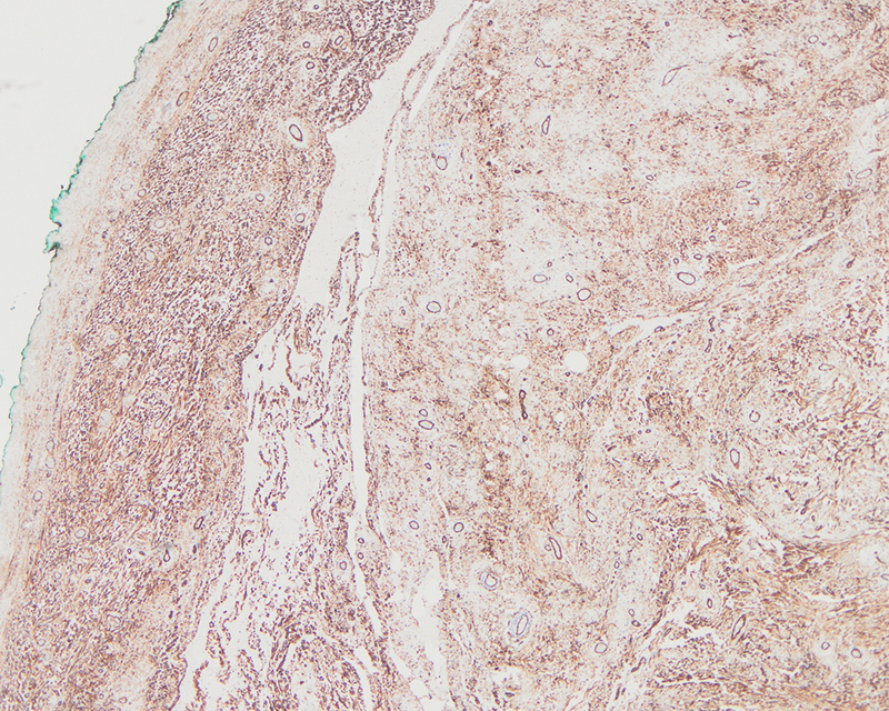
Complete surgical resection of the tumor revealed a well-circumscribed tan-white mass that measured up to 2 cm in greatest dimension. The cut surface was tan-white and firm without evidence of hemorrhage or necrosis. Microscopically, the lesion demonstrated prominent vasculature, pseudovascular spaces, and bland spindle cells in short fascicles with focal mild atypia (Figure 1 and 2). Intralesional fat was present (Figure 3). The lesion showed overall circumscription, with some irregular nodular extensions/prolongations of the lesion into the adjacent soft tissue resulting in the microscopic appearance of satellite lesions. Immunohistochemistry revealed strong CD34 positivity (Figure 4). Estrogen and progesterone receptors were weakly positive and desmin, SMA, and S-100 were negative.