Hard, Painless Breast Mass
By Claire Porterfield, BS and Huina Zhang, MD
Clinical History
75-year-old woman presented with a history of a 2 cm well-defined, hard, painless breast mass.
Recent History
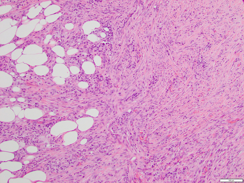
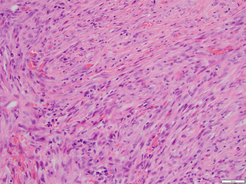
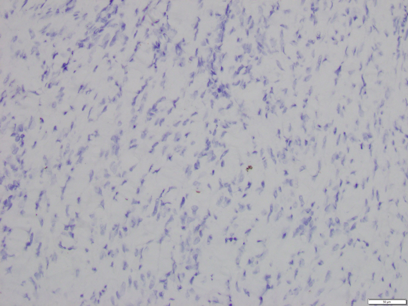
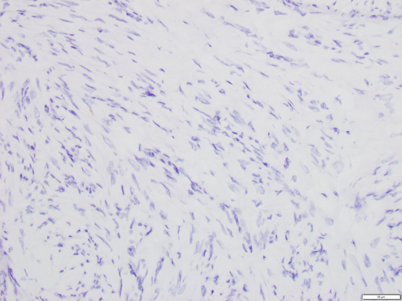
Limited patient history was available for review. The breast lesion consisted of a population of spindle cells with interspersed thick collagen bundles, focal lymphocytic aggregates and some extravasated erythrocytes (Fig 1). No overt cytologic atypia was observed and mitotic figures were infrequent (Fig 2).
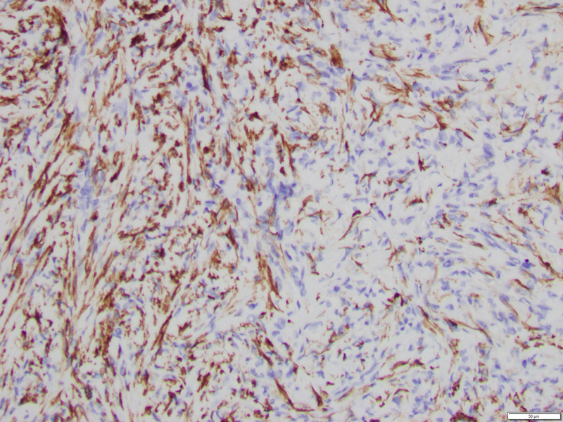
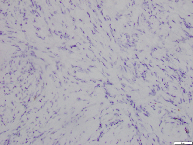
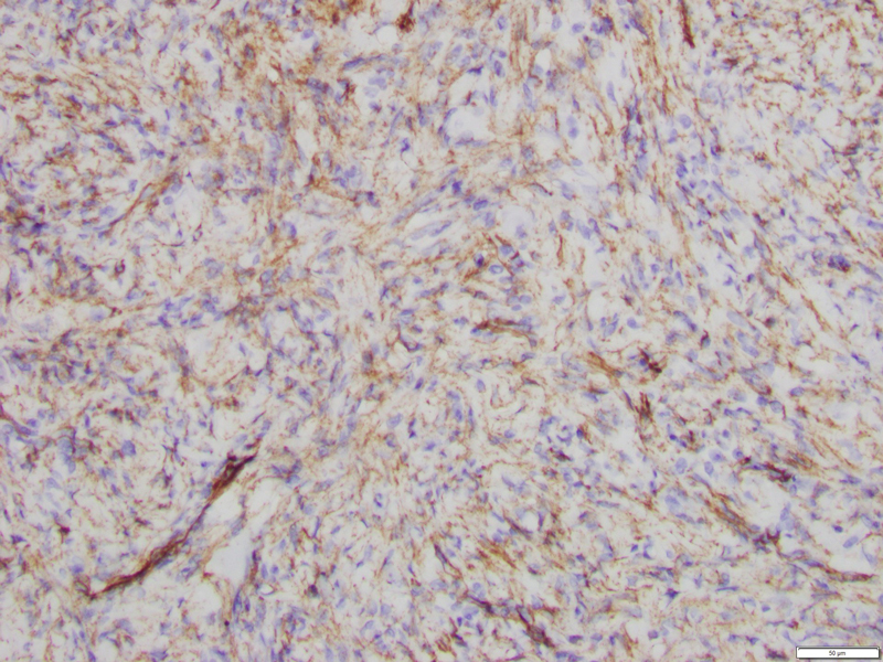
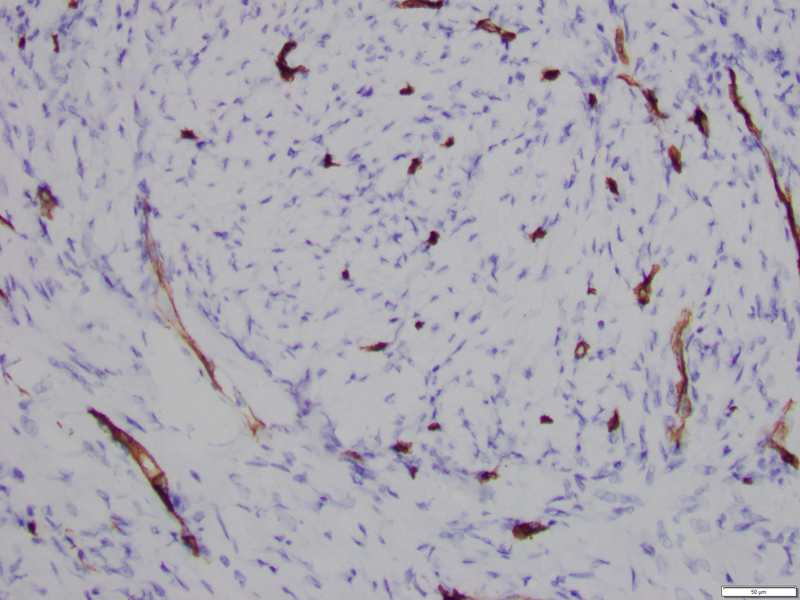
Immunohistochemical staining for smooth muscle actin (SMA) was positive, consistent with the presence of fibroblast cells (Fig 3). Whereas, stains for AE1/AE3, CK5, p63, desmin, beta catenin and CD34 (Fig 4-8, respectively) were all negative.