Case 78: Sigmoid Polyp on Screening Colonoscopy
Case 78: Sigmoid Polyp on Screening Colonoscopy
Roula Katerji, MD; Aaron R. Huber, D.O
Clinical History
An otherwise healthy 82- year-old patient presented for screening colonoscopy.
Past Medical History
The patient was asymptomatic with no prior personal or family history of colon cancer or inflammatory bowel disease.
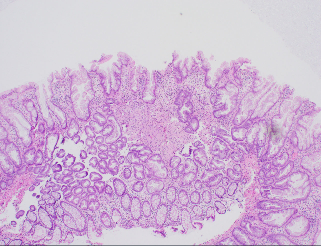
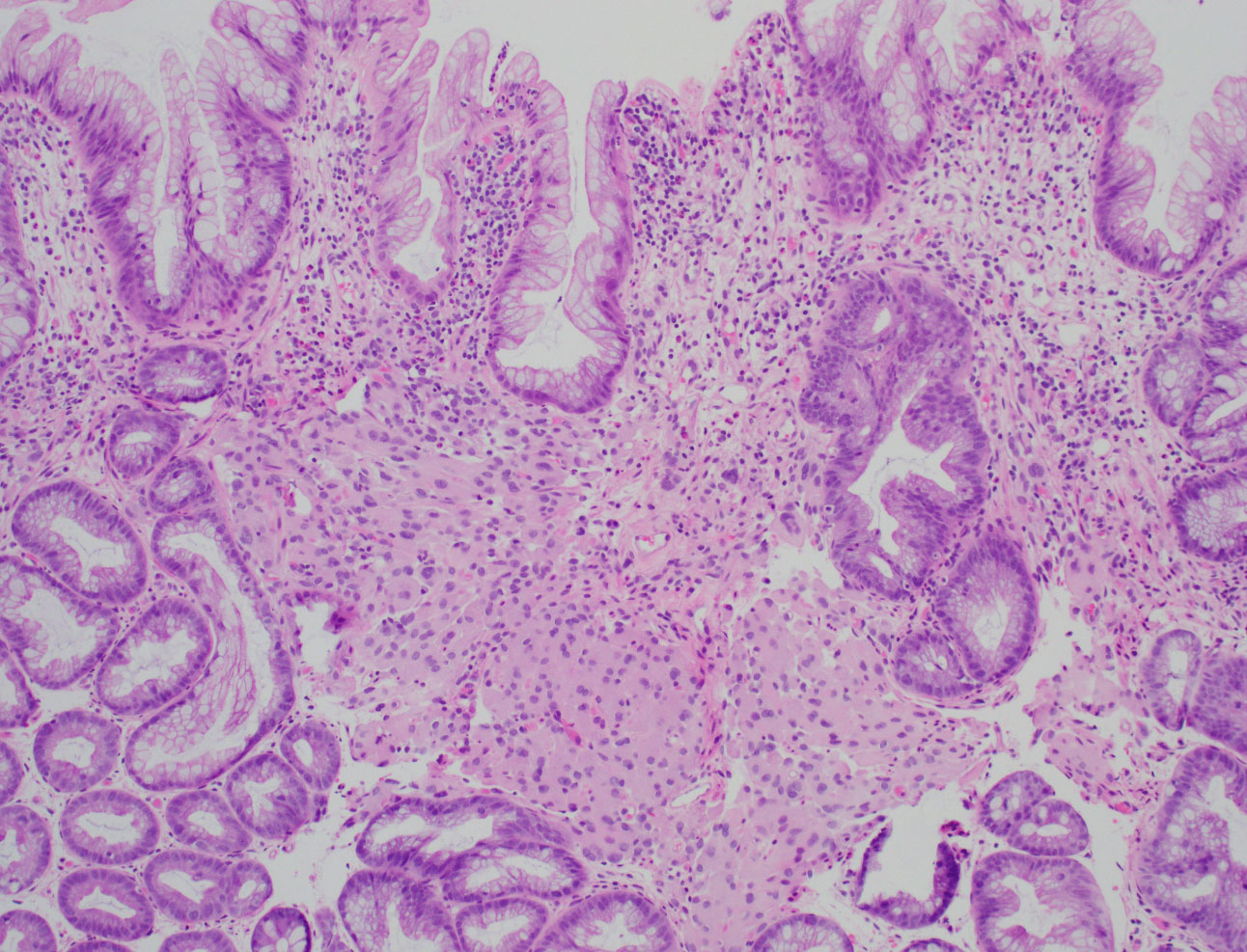
Pathology
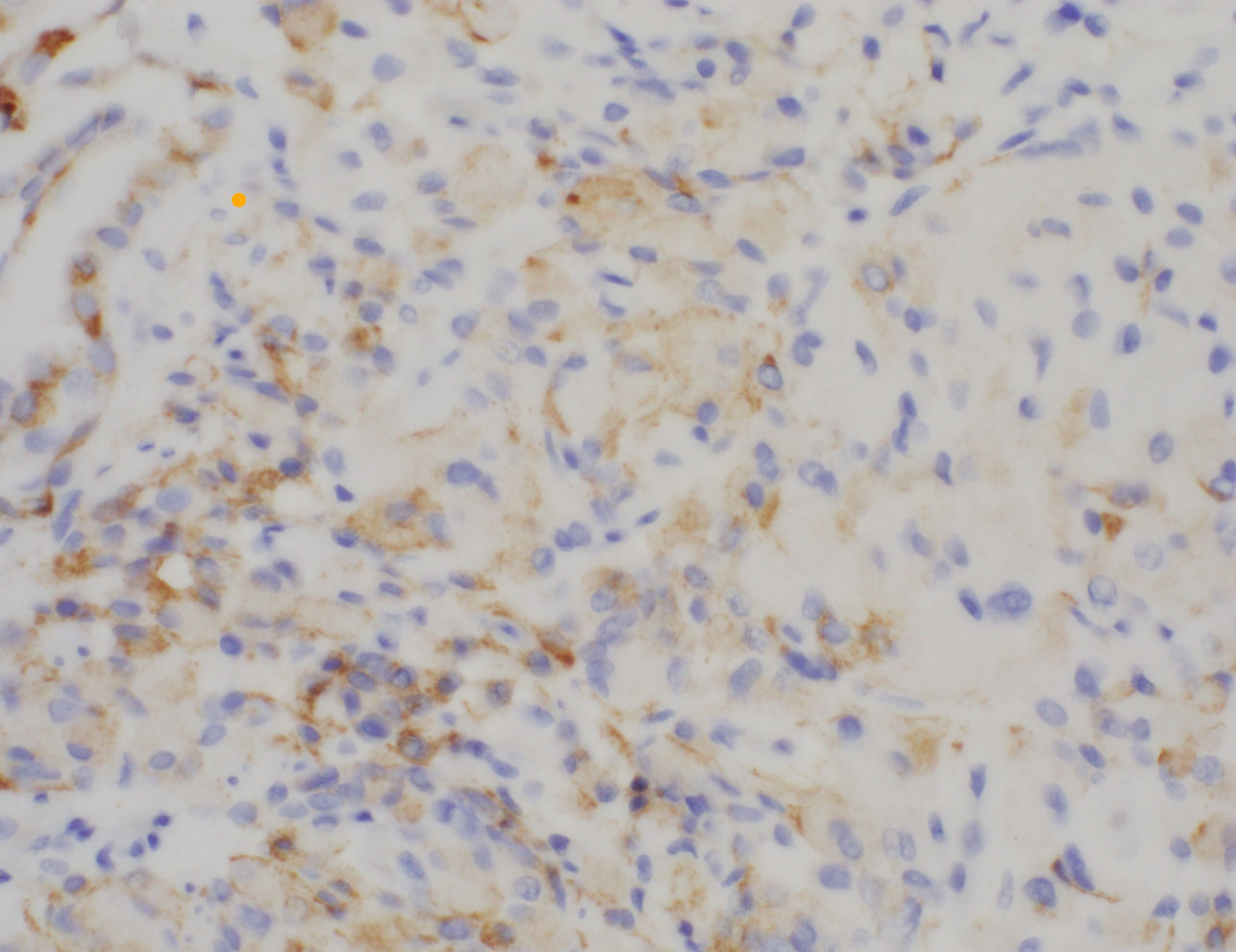
Screening colonoscopy demonstrated multiple polyps in the transverse colon and one diminutive (5 mm) polyp in the sigmoid colon. Histologically, the sigmoid polyp showed a hyperplastic polyp with an associated expansion of the lamina propria by a vaguely nodular proliferation of epithelioid cells. The epithelioid cells were arranged in small clusters with round nuclei, eosinophilic cytoplasm, and occasional nuclear grooves (Figures A-C). There was no cytologic atypia, mitotic activity, or necrosis. The epithelioid cells were positive for EMA (weak diffuse expression, figure D), while they were negative for pankeratin, GLUT1, S-100, SOX10, HMB45, DOG1, ERG, CD68, CD163, and SMA.