Imaging Services
St. James Hospital provides general imaging services 24 hours/day for inpatient, outpatient, and emergency needs.
Our Locations
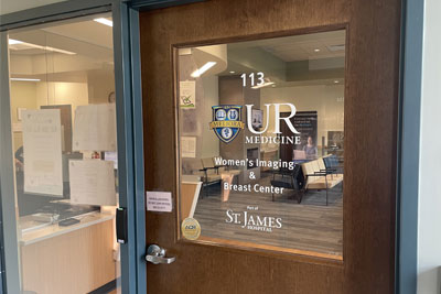
Women's Imaging & Breast Center
Part of St. James Hospital
7309 Seneca Road North
Suite 113, Entrance E
Hornell, NY 14843
Phone: (607) 385-3900
Appointment: (607) 247-2218
Hours vary by service,
please call to schedule an appointment.
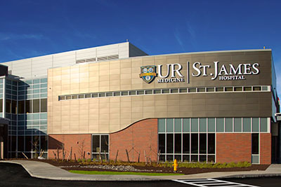
St. James Hospital
No appointment needed for X-Rays
Monday–Friday 7 a.m.–10 p.m.
7309 Seneca Road North
Hornell, NY 14843
Phone: (607) 247-2500
Appointment: (607) 247-2218
Hours vary by service,
please call to schedule an appointment.
Services
Women's Imaging & Breast Center
What are the Different Types of Imaging & Services?
Ultrasound
Ultrasound uses high-frequency sound waves to produce images. Doppler ultrasound may be used during the test. Doppler ultrasound is a technique that evaluates blood flow through a blood vessel. Typical ultrasound exams capture images from the abdomen (including during pregnancy), heart, gallbladder, breast, kidneys, arteries and veins.
- Breast Ultrasound: Breast ultrasound uses ultrasonography (sound waves) to image the breast. If an abnormality is seen on mammography or felt by physical exam, ultrasound is the best way to find out if the abnormality is solid, such as a benign fibroadenoma or cancer, or fluid-filled, such as a benign cyst. It cannot determine whether a solid lump is cancerous, nor can it detect calcifications. A breast biopsy is performed when a suspicious finding is discovered by mammography or ultrasound. An ultrasound biopsy is a minimally invasive procedure that uses ultrasound imaging to guide the needle, making it ideal for evaluating lumps or abnormalities that are visible on an ultrasound but not on a mammogram. These biopsies can be performed on site at our Women's Imaging & Breast Center.
- Pelvic and Obstetric Ultrasound: A pelvic ultrasound uses sounds waves to assess organs and structures within the female pelvis, including the uterus, cervix, vagina, fallopian tubes and ovaries. An obstetric ultrasound produces pictures of a baby (embryo or fetus), as well as the mother's uterus and ovaries. St. James has invested in new machines that include all the technological advances in obstetric ultrasound imaging. We are able to capture 3D images of your baby. Our technology does not use ionizing radiation, has no known harmful effects, and is the preferred method for monitoring pregnant women and their unborn babies. The Women's Imaging and Breast Center accommodates all OB patients.
Computed Tomography (CT)
We are equipped with state-of-the-art imaging equipment including a 64-slice CT scanner. CT scans are X-ray beams that rotate through narrow sections of the body. Many scans are taken in a short period of time and produce multiple images. A computer reconstructs the scans to produce two- and three-dimensional images. Typically, CT scans are captured of the head, chest, abdomen, and pelvis. The test can involve drinking contrast solution and/or IV contrast administration, which helps capture a better image. CT services are available on-site 24 hours/day.
DEXA Bone Density Scanning
DEXA (Dual-Energy X-ray Absorptiometry) Bone Density Scanning is a quick, non-invasive test that measures your bone mineral density using low-dose X-rays. It helps diagnose osteoporosis and assess your fracture risk by focusing on areas like the spine and hip. The test is painless, and the results guide your doctor in recommending treatments or lifestyle changes to strengthen your bones.
Magnetic Resonance Imaging (MRI)
Our in-house fixed MRI unit provides state-of-the-art imaging. MRI tests use a strong magnetic field, radio waves, and computers to create images. The MRI machine is cylindrical and creates a magnetic field around the patient. MRI is utilized in situations where organs or soft tissues are being studied; it does not use radiation. When examining the blood vessels, the MRI performs a specialized type of exam called an MRA (Magnetic Resonance Angiography). The MRI unit is a Phillips Ambition 1.5T. Interpretations are provided by the University of Rochester imaging team.
- Breast MRI: Our MRI machine has a special device called a dedicated breast coil to image the breasts. The MRI uses strong magnets to make very detailed, cross-sectional pictures of the body. MRI creates pictures of soft tissue parts of the body that would sometimes be hard to see using other imaging tests.
Unlike mammograms or breast ultrasound, breast MRI requires that you have a contrast dye injected into your vein (through an IV line) before the pictures are taken. This helps make any abnormal areas in the breast easier to see.
The MRI environment is calming and spacious and allows the patient to listen to music of their choice.
MyChart allows patients to view their imaging reports as soon as they are made available to the ordering provider.
Mammography
Mammography uses a dedicated digital X-ray machine to capture images of breast tissue. Regular mammograms are important in detecting breast cancer and other breast diseases. Mammograms should be performed annually per American Cancer Society guidelines. Mammography is often used in collaboration with ultrasound.
St. James offers the latest in 3D mammography, which provides clearer images especially for people with dense breast tissue. 3D mammography makes it easier for physicians to detect abnormalities, such as breast cancer and calcifications.
If necessary, biopsies, needle localizations, and cyst aspirations are done by recommendation of a radiologist. A stereotactic biopsy is a minimally invasive procedure that uses mammography to precisely locate the area of concern, guiding the needle to the exact spot for sampling. This method is especially useful for detecting small calcifications or masses not easily felt.
To make an appointment at our Women's Imaging & Breast Center, please call (607) 247-2218 or schedule your exam through MyChart.
Screening mammograms are covered by insurance and no co-pay is required. People who are not covered by insurance may contact Cancer Services Program at (607) 385-3933 or (877) 778-6857.
Echocardiogram
Echocardiograms use sound waves to show how blood flows through the heart and heart valves. Sensors attached to the chest and sometimes the legs check the heart rhythm during the test. The test can help providers diagnose heart conditions.
Pediatric Echocardiogram
A pediatric echocardiogram is a safe, noninvasive test that uses sound waves to create detailed images of a child’s heart. The exam helps providers see how the heart’s chambers, valves, and blood flow are functioning, allowing them to diagnose and monitor congenital heart defects, heart murmurs, and other cardiac conditions. The test is painless and performed using sensors placed on the chest.
The Ankle Brachial (ABI)
The Ankle Brachial Index (ABI) test is a simple, non-invasive procedure that compares the blood pressure in your ankle with the blood pressure in your arm. It helps to determine if you have peripheral artery disease (PAD), a condition where the arteries in your legs are narrowed or blocked, which can lead to pain and mobility issues. During the test, you will lie down, and a healthcare provider will use a blood pressure cuff and a Doppler ultrasound device to measure the pressures at both sites. The results will show how well blood is flowing to your limbs, guiding your doctor in diagnosing and managing PAD.
Radiology (X-ray)
X-ray images are typically captured to help diagnose disease and/or issues with the musculoskeletal system. An image is made by radiation in the form of X-rays passing through the body. The image is recorded digitally and available instantly. University of Rochester Medicine Radiologists then interpret the images and generate a report that is provided to your physician/provider. X-ray services are available 24 hours/day at the hospital.
- Fluoroscopy is a continuous X-ray beam is passed through the body part being examined. The beam is transmitted to a TV-like monitor so that the body part and its motion can be seen in detail. Services include joint injections, arthrograms, upper GI, esophagram, small bowel follow-through, barium enema, sniff test, modified barium swallows.
- C-Arm Machine, which are high-tech devices used by physicians to guide surgical instruments. The C-arms capture live images during surgical procedures. They provide high-resolution X-ray images immediately, which allow the physician to monitor progress at any point to make any immediate corrections. C-arms are used during pain management cases, general surgery and orthopedic procedures.