Model Systems and Techniques
Model Systems and Techniques
We employ several model systems and techniques in our laboratory including the following:
Model Systems
- Langendorff perfused mouse hearts (Picture 1)
- Isolation and culture of adult mouse cardiomyocytes
- Transverse aortic constriction induced mouse heart failure model
- Carotid artery partial ligation mouse model
- Femoral artery wire injury mouse model
- Abdominal aortic aneurysms mouse model
- Atherosclerosis mouse model
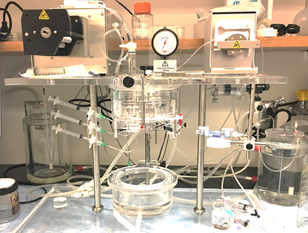
Picture 1. Langendorff heart perfusion system
Molecular, Biochemical, and Cell Biological Techniques
- Molecular cloning
- Western blotting
- Immunoprecipitation
- Immunostaining
- RT-PCR
- Transgenic gene expression
- Lentivirus/Adenovirus/Adeno-associated viruses-mediated gene delivery
Optical/Imaging Techniques
- Fluorescence energy resonance transfer (Picture 2)
- Confocal imaging
- Electron microscopy
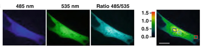
Picture 2. Representative image of primary rat cardiac fibroblasts transfected with cAMP FRET sensor Epac1-camps. Pseudo-colored fluorescence intensity is shown for the FRET donor, CFP (485 nm), the acceptor YFP (535 nm), corrected FRET ratio 485/535 nm.
Physiological Assays
- In vitro cardiomyocyte contraction (Picture 3)
- Ex vivo assessment of cardiac physiology
- Echocardiographic evaluation of cardiac function
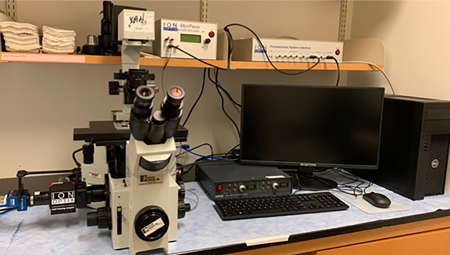
Picture 3. IonOptix Calcium and Contractility system for cardiomyocyte contractile function.