Projects
Circuit Tracing
 Neuroanatomy is the foundation of modern neuroscience. Recent technical innovations have made it possible to reconstruct large numbers of neurons in order to gather “high throughput” information about the different morphological cell types in the brain. One of the overarching goals of my research is to link structure and function in the visual system.
Learn more about Circuit Tracing
Functional Role of Corticogeniculate Feedback in Vision
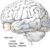
 In mammals, all conscious visual information traverses a path from the retina through the visual thalamus (called the lateral geniculate nucleus or LGN) to the primary visual cortex (V1). Accordingly, in just a handful of synapses and in about 50 milliseconds, information about a visual stimulus in the world can reach the cortex. In highly visual mammals such as humans, visual information traversing this path is segregated into three parallel processing streams. By separating out visual information that is most relevant for processing motion, form/acuity, and color, signals can be relayed in parallel for increased processing speed.
Learn more about Functional Role of Corticogeniculate Feedback in Vision
The Role of Visual Thalamus in Residual Vision Following Loss of Retinal or Cortical Inputs
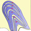 In mammals, the visual thalamus, the lateral geniculate nucleus (LGN), relays visual information from the retina to the primary visual cortex (V1) and also receives feedback from V1. Both retinal and cortical inputs are critical to processing visual information within the LGN. The role of the LGN in residual vision following loss of retinal or cortical inputs is not known. In particular, we want to explore how loss of inputs (retinal or cortical) alter the physiological responses of LGN neurons.
Visual Processing in Natural Settings
 Most of our knowledge about neuronal processing in the early visual pathways is based on studies in anesthetized or immobilized subjects in order to stabilize eye movements. These experimental setups introduce unnatural conditions that may limit the applicability of results to the real world.
Learn more about Visual Processing in Natural Settings
Neuronal Mechanisms of Attention
 The phrase “visual attention” captures a broad range of phenomena. Most people consider visual attention to be an active cognitive process. When we study visual attention in the laboratory, we mainly study covert visual spatial attention, or the allocation of attention to a particular region of visual space that is away from the center of gaze. We design visual attention tasks that involve shifts in the locus of covert spatial attention on different trials.