Mechanisms of Soft Tissue Fibrosis
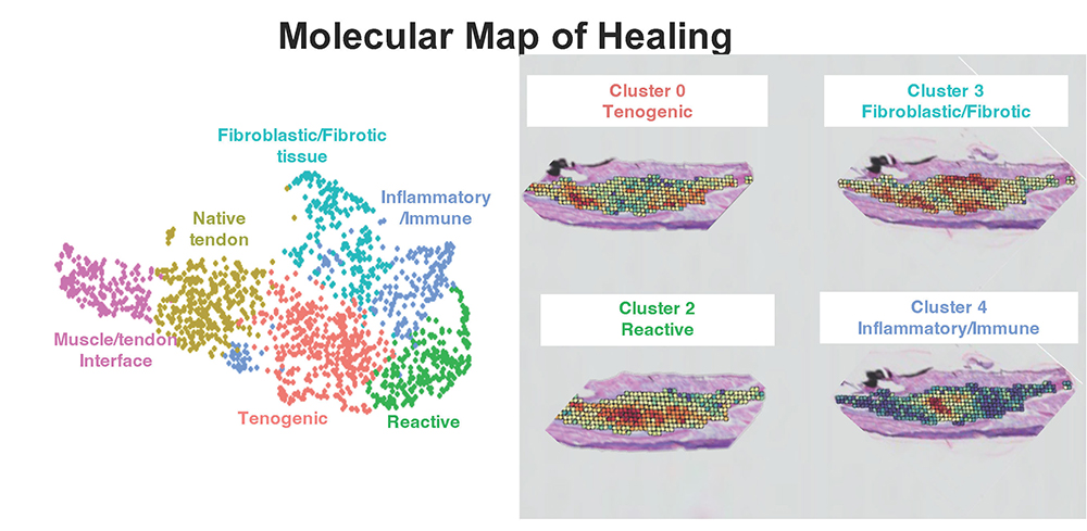
 Rather than regenerating native tendon tissue, tendons heal via a combination of an inner highly aligned ECM bridge and a surrounding shell of disorganized ECM scar tissue that is associated with the formation of peritendinous adhesions. As such, defining the spatially-distinct cellular and molecular programs that underpin these unique tissue features is critically for establishing therapeutic approaches that promote regenerative programs and blunt fibrotic processes. To address this key knowledge gap, we have conducted spatial transcriptomic profiling of the tendon healing process, and established key molecular drivers of different aspects of this process. On-going work is leveraging this large, comprehensive dataset to test new therapeutic candidates and to develop tendon-targeted drug delivery approaches.
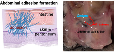
Abdominal adhesions result when scar tissue forms between the abdominal wall and visceral organs, such as the small intestine, in response to peritoneal tissue trauma during abdominal and pelvic surgery. Adhesion formation occurs in greater than 50% of cases, with the incidence rising to >95% in patients undergoing multiple surgeries. Abdominal adhesions are the number one cause of small bowel obstruction, can lead to infertility and chronic pain, and can complicate subsequent surgeries. Collectively, health care costs associated with abdominal adhesion-related complications exceed $5 billion annually in the United States. Currently, standard treatment options for abdominal adhesions are limited to laparoscopic or open surgical lysis. Thus, there is a substantial need to identify therapies to both prevent initial adhesion formation and to ameliorate adhesion persistence and recurrence.