News from the McGrath Lab
News from the McGrath Lab
Team Science Showcase: The People and Partnerships Moving Microplastics Research Forward
Monday, February 9, 2026
How a small project between a university lab and a city water system grew into a national model
Public concern about microplastics and health has grown sharply over the past several years. Questions like “Are plastic cutting boards safe?” and “Should I drink out of plastic water bottles?” are prevalent, but straightforward answers to these seemingly simple queries are not.
While new research on levels of microplastics found in the human body has recently raised flags for many, scientists at the University of Rochester have been asking questions, building teams, and uncovering how environmental exposures impact human health for decades. With the knowledge, partnerships, and systems they’ve put in place through the University’s Environmental Health Sciences Center (EHSC), now celebrating its 50th year of funding from the National Institutes of Health, they’re poised to provide answers.
“The EHSC was built on the idea that the biggest environmental health challenges require teams that cross disciplines, institutions, and communities,” said Paige Lawrence, PhD, chair of the department of Environmental Medicine at the University of Rochester School of Medicine & Dentistry. “Our microplastics work is a perfect example of how that model accelerates discovery. By bringing engineers, toxicologists, ecologists, and local partners together, we’re able to ask deeper questions and develop solutions that matter for people’s daily lives.”
The long-standing collaborative culture fostered by the EHSC led to the launch of the Lake Ontario MicroPlastics Center (LOMP) in April 2024. One of six federally funded Centers for Oceans and Human Health in the nation, LOMP marked the culmination of a decade of collaborative work among University of Rochester and Rochester Institute of Technology (RIT) researchers, community organizations, and local and state government to understand and reduce microplastic pollution in local waterways.
“Microplastics research in Rochester didn’t start with a single project—it started with relationships,” said Katrina Korfmacher, PhD, professor of Environmental Medicine at the University of Rochester School of Medicine & Dentistry. “For years, our faculty have worked alongside community and government partners who are deeply invested in water quality. LOMP is an extension of the EHSC’s long-standing commitment to doing science in partnership with communities.”
Korfmacher co-directs LOMP with Christy Tyler, PhD, professor in the Thomas H. Gosnell School of Life Sciences at the Rochester Institute of Technology. Today, their teams are working to understand how microplastics move through the Lake Ontario ecosystem and how they may affect human health under varied environmental conditions—research made possible by a discovery that connects back to the EHSC.
Read More: Team Science Showcase: The People and Partnerships Moving Microplastics Research ForwardRochester to advance research in biological imaging through new grant
Tuesday, February 23, 2021
A new multidisciplinary collaboration between the University of Rochester's departments of biology, biomedical engineering, and optics and the Goergen Institute for Data Science will establish an innovative microscopy resource on campus, allowing for cutting-edge scientific research in biological imaging.
Michael Welte, professor and chair of the Department of Biology, is the lead principal investigator of the project, which was awarded a $1.2 million grant from the Arnold and Mabel Beckman Foundation.
"The grant supports an endeavor at the intersection of optics, data science, and biomedical research, and the University of Rochester is very strong in these areas," Welte says. "The University has a highly collaborative culture, and the close proximity of our college and medical center makes Rochester ideally suited to lead advances in biological imaging."
The project will include developing and building a novel light-sheet microscope that employs freeform optical designs devised at Rochester. The microscope, which will be housed in a shared imaging facility in Goergen Hall and is expected to be online in 2022, enables three-dimensional imaging of complex cellular structures in living samples. Researchers and engineers will continually improve the microscope, and it will eventually become a resource for the entire campus research community.
In addition to funding the construction of the microscope and development of the data science component, the grant from the Arnold and Mabel Beckman Foundation supports three biological research projects:
- Anne Meyer, an associate professor of biology, will study how communities of bacteria interact and change inside lab-grown biofilms—three-dimensional structures that mimic how bacteria (including bacteria that cause disease) live in nature. Most microscopy is so slow that the bacteria may have already divided by the time the microscope is back at the same spot. The speed of light-sheet microscopy is expected to enable Meyer to see the bacteria in the 3-D space in real time.
- Dan Bergstralh, an assistant professor of biology, will study how the behavior of single cells generates the 3-D structure of animal organs. Using normal microscopy, researchers can either study single cells and their properties or the properties of tissues and organs. Light-sheet microscopy will allow Bergstralh to image both cells and organs at enough detail to understand how they influence each other.
- Jim McGrath, a professor of biomedical engineering, and Richard Waugh, professor of biomedical engineering and vice provost for research, will study how molecules and cells travel from the bloodstream into surrounding tissue. Traditional microscopy is insufficient to visualize critical aspects of this transport, but the new microscope will allow the researchers to look across a whole plane of many cells at the same time. It will also allow them to add drugs during imaging and immediately see how the sample reacts.
Awad, McGrath and Miller receive a $4M NCATS/NIAMS Clinical Trials on a Chip grant to study tendon inflammation and fibrosis
Tuesday, September 29, 2020
Clinical Trials on a Chip researchers plan to build and test common and rare disease models to help improve the clinical trial process.
Approximately 85% of late-stage clinical trials of candidate drugs fail because of drug safety problems or ineffectiveness, despite promising preclinical test results. To help improve the design and implementation of clinical trials, the National Institutes of Health has awarded 10 grants to support researchers' efforts in using tiny, bioengineered models of human tissues and organ systems to study diseases and test drugs. One major goal of the funded projects is to develop ways to better predict which patients are most likely to benefit from an investigational therapy prior to initiating clinical trials.
The awards total more than $6.9 million in the first year, and approximately $35.5 million over five years, pending available funds. They are administered through a new program, Clinical Trials on a Chip, which is led by NIH's National Center for Advancing Translational Sciences (NCATS) in conjunction with several other NIH Institutes and Centers, including the National Cancer Institute, the National Institute of Child Health and Human Development, and the National Institute of Arthritis and Musculoskeletal and Skin Diseases.
Tissue chips, or organs-on-chips, are 3-D platforms engineered to support living human tissues and cells and mimic complex biological functions of organs and systems. Tissue chips are currently being developed for drug safety and toxicity testing and disease modeling research, including on the International Space Station. Clinical Trials on a Chip is one of several initiatives that are a part of the NCATS-led Tissue Chip for Drug Screening program, which was started in 2012 to address the major gaps in the drug development process.
Rochester researchers pursue quick ways to detect COVID-19—and better understand it
Tuesday, April 21, 2020
University scientists are adapting existing research to develop tests to detect and improve our understanding of COVID-19. Examples include projects led by Martin Zand, senior associate dean for clinical research at the Medical Center; Benjamin Miller, a professor of dermatology and biomedical engineering; and James McGrath, a professor of biomedical engineering.
Zand is working on the finger-stick test, which uses patented technology that detects immunity to more than 50 strains of flu. The test comes in an easy-to-mail kit similar to those that test blood sugar for diabetes. "We're hoping this could make COVID-19 vaccine trials faster and more convenient for those who volunteer for them," says Zand.
Miller's lab hopes to find the virus with optics at the nanoscale. The lab is developing tiny sensor chips that use coronavirus proteins to "very quickly" detect the presences of immunoglobulin G and M antibodies that humans develop within two days of exposure to the virus. "The problem right now is actually getting patient samples," says Miller. "Meanwhile we are optimizing our assays with 'normal' serum samples doped with coronavirus antibodies—basically making artificial patient samples."
McGrath is using ultrathin membranes—less than 200 nanometers thick—to determine whether individuals have been infected with COVID-19. He can apply the membranes as a sensor and as a platform for discovering pathogenic mechanisms. McGrath is eyeing an inexpensive device similar to a pregnancy test that could be used in low-resource communities around the world.
"It will likely take more than a year to develop a vaccine, so COVID-19 is going to be with us for some time," says McGrath. "If we move quickly but deliberately, I think the device could be ready in time to help with the current pandemic."
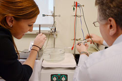
Detecting microplastics first step in assessing environmental harm
Thursday, January 9, 2020
Amid growing alarm over the plastic that pollutes our environment, biomedical and optics researchers at the University of Rochester are working to better understand the prevalence of microplastics in drinking water and their potential impacts on human health.
They are collaborating with SiMPore, a company that uses nanomembrane technology initially developed at the University, to devise ways to quickly filter and identify particles of plastic 5 mm or smaller in drinking water samples. They will then test the ability of these particles to cross a microscale barrier that simulates the lining of a human intestine.
"We want to see to what extent the particulates that you consume in your drinking water can pass through your gut and into your other organs," says Greg Madejski, a postdoctoral fellow in the laboratory of James McGrath, professor of biomedical engineering. Madejski is coordinating the research with the lab of Wayne Knox, professor of optics. Both McGrath and Knox are affiliated with the Materials Science Program.
Microplastics are used as ingredients in cigarette filters, textile fibers, and cleaning or personal care products. Others result when larger plastic items are worn down by sun, wind, and waves. They can be found on mountaintops and at the bottom of the oceans; in the air we breathe and in the water we drink. Exactly how many microplastics are absorbed by humans, and how much harm it is causing them has been hard to assess because the particles— below 100 microns—are so small and difficult to detect.
"These are particles that you couldn't pick up with tweezers; that you can't even see with the naked eye," Madejski says. They elude the "traditional method of skimming the surface of water with a plankton net and collecting everything," he says.
Instead the researchers will filter water through sheets of silicon nitride a hundred times thinner than the diameter of a human hair. These SiMPore nanomembranes, based on prototypes initially created in the McGrath lab, have micron-sized slits in them. "That allows us to catch micron-sized debris," Madejski says. "And because the sheets are so thin, you can filter a significant amount of water through them without a lot of pressure."
Professor Jim McGrath receives HSCCI Grant
Monday, July 30, 2018
Professor Jim McGrath has received a Health Sciences Center for Computational Innovation (HSCCI) Grant for his project, "Database of Fluid Flow in Nanomembrane-based Microdevices." This project seeks to build a database of computational 3D flow profiles for microfluidic devices featuring the laboratory's silicon nanomembranes. Nanomembranes are ultrathin porous membranes with orders of magnitude higher permeability than conventional membranes. A two-channel, microfluidic device with a 5.4 mm square membrane 'chip' separating the channels has become a standard format for multiple projects in the McGrath laboratory. Specifically, the device format is used for: 1) benchtop evaluations of toxin clearance in hemodialysis, 2) capture of exosomes from raw biofluids in tangential flow, 3) a 'tissue-chip' mimetic of the microvasculature. Future microfluidic applications of this device include: 1) enhancement of DNA detection by a novel dual membrane system, and 2) an electrodialysis system for desalinization.
The Health Sciences Center for Computational Innovation (HSCCI) facilitates access to high-performance computational resources for biomedical research. HSCCI is the result of a partnership between the University of Rochester, IBM, and New York State. The University seeks to develop the center further through corporate partnerships, institutional support, federal research grants, and New York State programs.
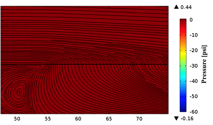
2D COMSOL simulation of flow near membrane (yellow). Flow is left to right on top with some transmembrane flow. Fluid streamlines are shown as black lines.
McGrath Lab graduate student’s accidental exhale leads to improved DNA detector
Thursday, December 7, 2017
Doctoral student Greg Madejski's illustration of the layers comprising his new DNA detection device. (University of Rochester illustration / Greg Madejski)
Greg Madejski held his breath as he looked into the microscope, trying to weld two fingernail-sized chips together: a tiny chip containing a nanofilter on top of another chip with a DNA sensor.
It was frustrating work. The chips weren't making good contact with each other. Madejski gently poked at the chips, then peered over the top of the microscope.
And exhaled.
The sudden waft of warm air swept over the nanofilter, transferring it to the sensor—right on target. The "accident" led Madejski to an important insight: the water vapor in his breath had condensed on the device, causing the nanofilter to adhere ever so neatly to the sensor.
"It was like a really high-tech temporary tattoo that I created by accident; lick and stick!" says the PhD student in the lab of James McGrath, a professor of biomedical engineering at the University of Rochester.
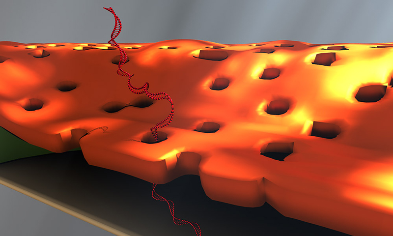
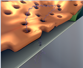
And that's how water vapor became integral to the development and design of a novel device for detecting DNA biomarkers affiliated with disease. Created by McGrath's lab in collaboration with Professor Vincent Tabard-Cossa and graduate student Kyle Briggs at the University of Ottawa, the device is described in an article published online at Nano Letters. The article, and an image from Madejski's homemade animation of the device in operation, will be highlighted on the cover of the February 2018 print issue.
This animation shows, as graduate student Greg Madejski explains, the "thin films of water, seen as rainbow colors, swelling and shrinking the space between the prefilter and the nanopore as its exposed to additional water vapor.
'A remarkable structure'
The device is comprised of three ultrathin layers:
- a nanoporous silicon nitride membrane which serves as a prefilter.
- a biosensor membrane with a single nanopore.
- a spacer layer that separates these by only 200 nm.
The arrangement creates a nanocavity filled with less than a femtoliter of fluid—or about a million times smaller than the smallest raindrops.
During operation, the device uses an electric field to lure a strand of DNA to enter one of the pores of the prefilter and then pass through the nanocavity to reach the pore of the underlying sensor membrane. This triggers changes in the device's electrical current that can be detected and analyzed. The fact that DNA must elongate itself in a consistent way to pass through the two-membrane combination improves the precision and reproducibility of detection.
"This is a remarkable structure," says McGrath. "We've built an integrated system with a highly porous filter within molecular reach of a sensor. I think there are many sensors, particularly those that hunt for biomarkers in raw biological fluids, that would benefit from filtering away unwanted molecules immediately upstream of the detector."
The method of fabrication instantly wets the nanocavity, which is often difficult at the nanoscale. The device contains dozens of these nanocavities, which may eventually increase the amount of material that can be screened by enabling parallelized biomarker detection.
Solving problems that others need solved
Tabard-Cossa's lab uses solid-state nanopore devices to find new ways to manipulate and characterize single molecules. His lab was interested in finding new materials that could be used for biomarker detection. The prefilter in the new device addresses a problem with other silicon nanopore detectors: They are more likely to clog than alternative devices that use that biological pores for sensing. Biological membranes, on the other hand, are less stable than solid state nanopores, McGrath noted.
"We love to apply our membrane technologies to solve problems that others need solved. This is a very nice example.," McGrath says.
McGrath is co-founder of SiMPore, a University-based startup that develops highly portable, chip-based devices that incorporate silicon membranes for a variety of applications, from biological sensing to dialysis.
"I think we're going to realize the practical advantages of this technology in the near term," he says. A second generation of the new device, developed at SiMPore, incorporates the prefilter right on the chips during manufacturing at the wafer scale, "so there's nobody breathing on it anymore," he notes. "It's actually all built as one unit and should make future studies very easy. That's a credit to the ingenuity at SiMPore and quite a legacy for Greg."
URMC Awarded Nearly $6 Million to Study Deadly Bone Infections
Monday, November 6, 2017
Bone infection, while relatively rare, can be debilitating and potentially fatal. In recent years, researchers in the Center for Musculoskeletal Research at the University of Rochester Medical Center have made several discoveries that position them to advance new treatments and possible cures for bone infections. Now, a nearly $6 million, 5 year award from the National Institute of Arthritis and Musculoskeletal and Skin Disease at the National Institutes of Health, will allow the group to create a new multidisciplinary research program devoted to studying bone infections.
The CMSR has been among the top five NIH-funded orthopaedic research centers in the nation for over ten years, and Edward Schwarz, Ph.D., Burton Professor of Orthopaedics and director of the CMSR, has been the top NIH-funded orthopaedic researcher in the nation three years running. This new grant, awarded to Schwarz and throng of researchers from across the University of Rochester and beyond, brings the center's total forecasted earnings for 2017 to $28 million.
Of the millions of Americans who have joint replacement surgeries each year, less than five percent come away with an infection. But this minority of patients must endure a long and difficult road to recovery, if they recover at all. The vast majority of these infections are caused by a bacteria called Staphylococcus aureus, including the dreaded methicillin-resistant strain (MRSA), which causes sepsis and death in 13 percent of infected patients.
Patients who survive these infections face multiple surgeries to remove infected tissue, months of strong antibiotic treatments, and a high likelihood of re-infection. For a long time, researchers have been working to understand how this bacteria evades treatment and Schwarz believes he has figured out.
Together with Karen Bentley, director of the Electron Microscopy Core at URMC, Schwarz showed that the bacteria can crawl deep into tiny channels in bones, possibly taking shelter there and later emerging to re-establish an infection. Though S. aureus was originally thought to be incapable of movement, Bentley and Schwarz, in collaboration with James McGrath, Ph.D., professor of Biomedical Engineering at URMC, and his spin-off company, SiMPore Inc., showed that this bacteria can migrate through tiny pores in membranes in the lab.
This new grant will allow Schwarz and Hani A. Awad, Ph.D., professor of Biomedical Engineering and Orthopaedics in the CMSR, to investigate exactly how S. aureus gets into bone and develop new treatments that target those mechanisms. Microbiologists Steven Gill, Ph.D., and Paul Dunman, Ph.D., in the Department of Microbiology and Immunology, will help the team develop new antibiotics to inhibit bone infection, which will be 3D printed into spacers that replace infected joint implants. Delivering the antibiotic at the site of infection may save patients' limbs and lives.
Schwarz has also been working to understand what makes certain patients more susceptible to S. aureus infections than others, including why some patients recover relatively easily, while others die.
"Death following surgical site infection is not random," said Schwarz. "By studying patient immune responses to this bacteria, we might be able to predict who will be fine and who will need extra medical attention."
S. aureus can also become resistant to antibiotics, making it extremely deadly and difficult to eradicate. Better understanding patients' immune reactions to the bacteria may provide new approaches to defeating it.
In an international study of more than 300 patients with infected total joint replacements, Schwarz and his team including John Daiss, Ph.D., and Chao Xie, M.D., in the CMSR, found that patients fared well if their immune systems attacked a certain S. aureus protein, and poorly if they attacked another. Patients who produced antibodies attacking autolysin, a protein important for cell division, were protected. Those who produced antibodies against a family of iron sensing determinant (Isd) proteins, which help S. aureus sap nutrients from its host, were more likely to experience sepsis and even die.
It is unclear why antibodies that attack Isd proteins are bad for patients, and Schwarz is determined to use this new funding to figure it out. He will also analyze the full complement of antibodies produced by patients infected with several types of staph bacteria to see if there are more good- and bad-cop antibodies that could help inform new treatments.
The Clinical Research Core of this program will be run by Stephen L. Kates, M.D., at Virginia Commonwealth University.
Professor McGrath receives NIH funding for collaborative research project with the University of Ottawa
Tuesday, May 30, 2017
Nanoporous Nitride nanomembrane used as
a pre-filter for a DNA biosensor.
Professor Jim McGrath has received National Institutes of Health funding (R21) for his research project, “Solid-State Nanopores integrated with Nanoporous Membranes for enhanced Single-Molecule Counting of low-abundance Biomarkers,” in collaboration with the University of Ottawa.
This project aims to create robust biosensors by combining a University of Ottawa technology for the electrical detection of individual DNA molecules with University of Rochester’s nanomembrane technology. The porous nanomembranes serve as protective filters for the DNA sensors and prevent large molecules and debris from reaching the biosensor while still allowing the unobstructed passage of DNA. The technology will be used in a strategy in which designer DNA ‘barcodes’ serve as amplified surrogates for low abundance biomarkers present in biological fluids.
Professor McGrath receives NSF funding for research collaboration with RIT and SiMPore, Inc.
Friday, May 26, 2017

Figure: Concept of a compact extracorporeal dialysis
system enabled by silicon nanomembranes
A collaborative project including Rochester Institute of Technology, SiMPore, Inc., and BME Professor Jim McGrath titled, “Development of Ultrathin Nanomembranes for Home-based Hemodialysis,” has received National Science Foundation funding. This collaboration between University of Rochester, RIT, and SiMPore Inc. continues the development large area silicon nanomembranes for wearable hemodialysis. The high efficiency of the ultrathin nanomembranes enables new form factors for hemodialysis that can dramatically improve both quality of life and health outcomes for those with end stage renal failure.
BME PhD candidate Kilean Lucas takes second place at the Hajim School's Art of Science Competition
Saturday, May 6, 2017

Blood Cells by Scanning Electron Microscope

“You see some phenomenal things under a scanning electron microscope,” says Kilean Lucas.
Such as the image above, which the PhD student took of a single human red blood cell, captured among several white blood cells on a silicon nanomembrane developed in the lab of Lucas’ advisor, James McGrath, professor of biomedical engineering.
Lucas is studying how the ability of these nanomembranes to separate and filter out particles that differ by mere microns in size could lead to medical breakthroughs. For example, immature red blood cells separated from mature red blood cells could be harvested and seeded into bioreactors as a new way to replenish blood banks.
Or all the white blood cells could be filtered out from a patient’s blood sample. There are many forms of white blood cells, each responding in different ways to different threats. The proportion of each type of white blood cell in a given sample could be used to give a rough diagnosis of the body’s response to disease.
Lucas said it was not easy deciding which of several images from his research to submit to the Art of Science competition. “This one had a very good balance of being far enough out where we could see multiple types of cells, but not so close that we’re only looking at one or two,” Lucas says. “What I tried to do with the pseudo coloring is to emphasize that variety.”
He hopes this image will convey to people how “stunning it is, in and of itself, that we’re able to capture all of these cells out of blood, a very complex solution” – in ways that could change peoples’ lives.
Read more about this year's Art of Science Competition here.
Next stop for Falling Walls winner: Berlin
Friday, April 21, 2017
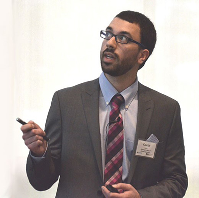
Biomedical engineering doctoral student Kilean Lucas delivers his presentation
at the annual Falling Walls competition. (University photo / Bob Marcotte)
Kilean Lucas admits to being uneasy about trying to summarize his research in three minutes, with only three slides.
You wouldn’t have known it yesterday, when Lucas, a PhD student in biomedical engineering, placed first at the University’s Falling Walls competition.
Lucas won $500 and an all-expenses paid trip to represent the University at the international Falling Walls competition in Berlin this fall.
Lucas described how the silicon nanomembranes developed in the lab of his advisor, James McGrath, could be used to filter out telltale exosomes (small, cell-derived vesicles) from the blood to provide early detection of cancer.
“There’s a lot of weight off my shoulders,” he said afterwards. “I was very nervous about coming to this and being able to communicate my project clearly in three minutes. That’s the hardest part of this.”
So how did he prepare?
With a big assist from his mentor and colleagues.
“I came up with the slides with help of professor McGrath,” Lucas said. Then I came up with a script that I tried out on my lab mates.”
Their feedback helped Lucas through four revisions of the script.
”Then I got together a group of people including my advisor and random people around the department and did a dry run. They gave me a lot of feedback,” Lucas said. In fact, he added, laughing, “ They tore my script apart. I completely reworked it yesterday.”
Sixteen competitors presented in front of a faculty panel that included jury chairman Richard Waugh, interim dean of faculty and associate vice president for research; Kim Arcoleo, associate dean for research at the School of Nursing; Dirk Bohmann, senior associate dean for basic research at the School of Medicine and Dentistry (SMD); Stephen Dewhurst, vice dean for research at SMD; Wendi Heinzelman, dean of the Hajim School of Engineering and Applied Sciences, and Joan Saab, chair of art and art history.
Runner up Kevin Mazurek, a post doctoral fellow in neurology, described a neurorehabilitative approach to help victims of stroke or other brain injury regain the ability to perform daily tasks.
Neurosurgery resident Jonathan Stone, the third place winner, described how patient-specific models of simulated tissue could help surgeons practice difficult procedures ahead of time.
The Falling Walls competition commemorates the fall of the Berlin Wall by giving young entrepreneurs and inventors from around the world the opportunity to express ideas about how to “break down the walls” hindering progress in dealing with challenges confronting science and society.
Lucas’ three-minute presentation:

Today I am going to talk to you about breaking the wall of cancer diagnostics.
Over the course of our lifetime, we have a 40 percent chance of developing cancer, and a 20 percent chance of dying of cancer. Late stage cancers present the highest risk of death as they are like a hydra.
Removing one tumor, or one head, does not guarantee that we kill the whole beast. In fact early diagnosis of cancer can save the lives of over 4 million people around the world each year.
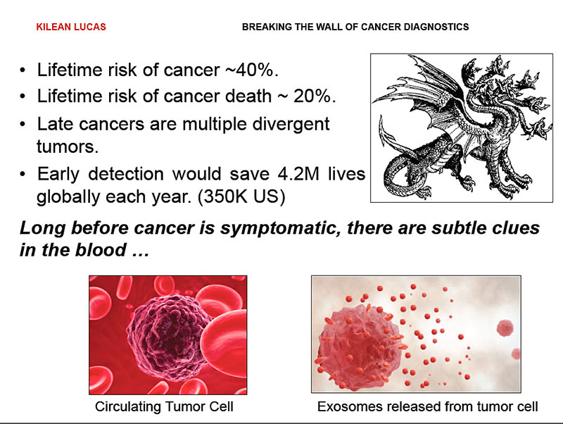
Conveniently, long before cancer presents outward symptoms, it releases subtle clues into the bloodstream in the form of circulating tumor cells and exosomes –50 nanometer vesicles that are basically breadcrumbs of information,
These exosomes contain micro RNA and proteins that are unique to the cells of origin, and provide us with a fingerprint of all the cells present in our body.
While they have all the information we need for diagnosis, searching for them is like searching for needle in haystack. Blood is a multicomponent system, and this makes it very difficult to easily search for these breadcrumbs.
However, what’s the best what to look for a needle in a haystack?
We need to filter out the hay.
For blood, we have developed exactly the filters we need to do this.
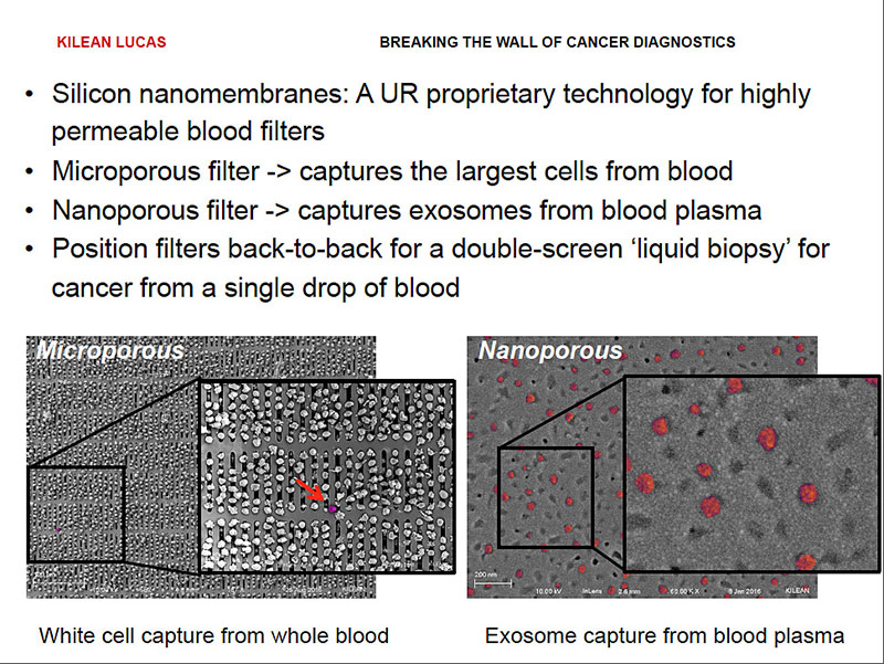
Silicon nanomembranes are one-of-a kind-technology that has been developed right here at the University of Rochester. We have the ability to uniquely tune their pores for highly specific size cutoffs.
This allows us to take membranes with pores that are micrometers in size to completely remove all the cells from blood, including circulating tumor cells, leaving us with only the exosome containing plasma. We can then take that plasma and pass it over second membrane with nanometer sized pores, filtering out remaining proteins and capturing the exosomes.
By pairing these membranes back to back, we can incorporate them in a single device that we can put in front of a routine blood test, giving us an unobtrusive liquid biopsy for breaking the walls of early cancer diagnostics.
Professor McGrath receives Dean's Office PumpPrimer II Grant
Thursday, October 6, 2016
Professor Jim McGrath recently received a Dean's Office PumpPrimer II Grant for his research project titled, "Desalinization with Ultrathin Nafion Membranes.”
Project description: The World Economic Forum’s Global Risks Report has consistently ranked access to water as one of the most critical issues facing the planet. Arid and drought-strikes regions with ready access to sea water in California, Israel, India, Australia, and elsewhere are investing billions in the production of inefficient reverse osmosis (RO)-based desalinization plants to produce drinkable water. Increasingly, technologists are turning to nanotechnology as a means of reducing costs by fundamental changing the fundamental principles at work. This project will test a prediction that ultrathin (100 nm thick) Nafion® membranes have the potential for desalinization with orders-of-magnitude greater efficiency than conventional reverse-osmosis (RO). This prediction is based on unexpected findings of a rate of osmotic flux of pure water across ultrathin Nafion® membranes used as electroosmotic pumps.
Karl Smith places third in University’s Falling Walls Competition
Wednesday, May 18, 2016
Karl Smith, a PhD student in Biophysics and a member of the lab of James McGrath, Professor of Biomedical Engineering, won third place in the University of Rochester's Falling Walls Competition for describing his use of physics to make water behind a filter form a mixer vortex, reducing the difficulty of normal stirring when fluids stick to surfaces. A total of 19 presenters competed.
The competition is associated with the Falling Walls foundation, a non-profit organization that fosters discussions on research and innovation and promotes the latest scientific findings to society. The Rochester winner's idea will compete with others from around the world at the Falling Walls Lab Finale in November in Berlin. This event selects the participants for the annual Falling Walls Conference the following day: an international forum for science and innovation to commemorate the fall of the Berlin Wall. Speakers at the conference have included Angela Merkel, Chancellor of Germany; Nobel Prize winner Sir Paul Nurse; and young inventors from around the world. BBC London said it was where the "brightest minds on the planet" meet.
Last year's Falling Walls Lab Rochester winner, Ryan Trombetta, a BME PhD student in Dr. Awad's lab, finished 12th (out off a 100 finalists worldwide) in the Berlin competition for his description of using 3D printed bone grafts to treat osteomyelitis. See his presentation here.
From left to right, Solomon Abiola, Sara Nowacki and Karl Smith, the top three finishers at the Falling Walls Competition.
Grant Will Help Move UR Innovations From Bench-Top to Bedside
Tuesday, February 3, 2015
BME Professor Jim McGrath has received I-Corps funding for his project entitled Portable Hemodialysis
which aims to develop a portable hemodialysis system for acute renal replacement therapy that clears toxins at rates required for human treatments. The McGrath lab will develop a multichip dialysis prototype that clears urea (acute kidney failure) and ammonia (acute liver failure) from blood at a rate (10 mL/min) typical of standard dialysis machines.
The NSF I-Corps program gave us an opportunity to investigate the commercial viability of our ideas for wearable hemodialysis by talking to 100 potential ‘customers.’ These customers included patients, doctors, caregivers, dialysis center and hospital administrators, first responders, engineers, social workers and more. The experience transformed our understanding of hemodialysis, how it is administered, and where the technology needs actually are. We’ve used the I-Corps experience to write highly informed and focused grant proposals to the NIH that we hope will enable us to translate our technology and actually improve the life of patients with end stage renal failure
, said McGrath.
Professor Awad and Professor McGrath Inducted into AIMBE College of Fellows
Wednesday, January 14, 2015
Department of Biomedical Engineering Professors Dr. Hani Awad and Dr. James McGrath were recently inducted as American Institute for Medical and Biological Engineering (AIMBE) Fellows for their significant contributions to the biomedical engineering community.
AIMBE's College of Fellows includes around 1,500 individuals who have made significant contributions to the medical and biological engineering community whether in academia, industry, or government and their contributions to research, industry practice, and education have transformed the world.
SiMPore and Micropen Announced as Winners of CEIS 2013-2014 STAR Program
Monday, January 13, 2014
The Center for Emerging & Innovative Sciences (CEIS) has announced SiMPore and Micropen Technologies as the winners of the 2013-2014 Short Term Applied Research (STAR) program. The STAR program focuses on New York State small businesses to address and solve time critical science and business problems.
SiMPore is a Rochester, N.Y.–based nanotechnology company co-founded by James McGrath, Associate Professor of Biomedical Engineering and Graduate Program Director of Biomedical Engineering at the University of Rochester. SiMPore designs and produces membranes and membrane-enabled products based on its unique patent-pending platform technology—the NanoBarrier™ ultrathin nanoporous silicon membrane. The NanoBarrier™ membrane is the world’s first membrane to offer both tunable nanometer-scale thickness and pore size. SiMPore is developing products that take advantage of these one-of-a-kind features, including filters for separating and concentrating biological molecules and nanoparticles, cell culture substrates for growing cells, and electron microscopy grids for preparing and imaging samples at the nanoscale. For more information please visit SiMPore.
Micropen Technologies is a design, development, and manufacturing resource and partner to electronics companies and medical device companies in the specialized technology of applying functional materials to surfaces.
Micropen Technologies has collaborated with the University of Rochester for more than a year on medical balloons with ablation electrodes and temperature sensors that can precisely apply energy to deactivate or destroy targeted nerves. In particular, denervation of renal nerves holds great promise in treating patients with drug-resistant hypertension. The work started as a Senior Design Project in the Biomedical Engineering Department and has continued at the Center for Medical Technology & Innovation. The goal is to develop a universal printed balloon solution for denervation therapies applied anywhere in the body. For more information please visit Micropen Technologies.
Super-Thin Membranes Clear the Way for Chip-Sized Pumps
Monday, October 28, 2013
A microfluidic bioreactor consists
of two chambers separated by a nanoporous silicon membrane.
The ability to shrink laboratory-scale processes to automated chip-sized systems would revolutionize biotechnology and medicine. For example, inexpensive and highly portable devices that process blood samples to detect biological agents such as anthrax are needed by the U.S. military and for homeland security efforts. One of the challenges of lab-on-a-chip
technology is the need for miniaturized pumps to move solutions through micro-channels. Electroosmotic pumps (EOPs), devices in which fluids appear to magically move through porous media in the presence of an electric field, are ideal because they can be readily miniaturized. EOPs, however, require bulky, external power sources, which defeats the concept of portability. But a super-thin silicon membrane developed at the University of Rochester could now make it possible to drastically shrink the power source, paving the way for diagnostic devices the size of a credit card.
Up until now, electroosmotic pumps have had to operate at a very high voltage - about 10 kilovolts,
said James McGrath, associate professor of biomedical engineering. Our device works in the range of one-quarter of a volt, which means it can be integrated into devices and powered with small batteries.
McGrath's research paper is being published this week by the journal Proceedings of the National Academy of Sciences.
BME Alumni Jess Snyder leads her team to International Victory!
Wednesday, May 22, 2013
The University of Rochester women's team beat out the others, completing the 3.5-mile course of Corporate Challenge Championship in a combined 1 hour, 24 minutes and 41 seconds. UR's Jessica Snyder (running the course in 20 minutes, 19 seconds) led Sarah Loerch, Kristina Maletz, and Christina deVries across the finish line.
It was the first time Rochester hosted the international championships; 10,921 runners registered for the regular race, which took place at the same time and venue as the championships.
UR, RIT Researchers Face New Pressure to Commercialize Work
Thursday, January 3, 2013
When University of Rochester scientist James McGrath started his career, doing research that would lead to marketable products was not a priority.
But the landscape has changed dramatically. Government funding for research has been stagnant for several years. Public and private grants now come with greater demands for results that can help drive profits and economic development.
Some venture capital firms are investing less in small early-stage projects. Pharmaceutical companies and manufacturers have switched from in-house research to working with universities and other institutions. Partnerships between university researchers and industry have grown.
Researchers Working on Dialysis Machine Small Enough to Hold in Your Hand
Wednesday, January 2, 2013
Imagine a dialysis machine small enough that a patient could wear it. A super-thin filtering material may allow researchers at the University of Rochester to revolutionize dialysis for patients with kidney disease. Jim McGrath, an associate professor of biomedical engineering at the University of Rochester, says the thinner the membrane that blood passes through, the more efficient its filtering capacity.
McGrath says the material they're working with filters blood more efficiently, and could end up in a much smaller device that could fit on an arm band. We can basically replace the experience of going to a dialysis center three times a week with nightly dialysis at home with a device that's about the size of a cell phone and achieve the same sort of clearance level. This is actually like a clinic on a chip,
he said.
Super-Thin Membranes Used in Lab-on-a-Chip Diagnostic Devices
Monday, November 12, 2012
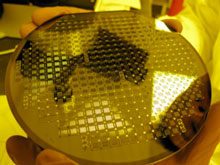
4" wafer with 160 membranes. (Photo by SiMPore Inc.)
Nano-porous silicon membranes developed at the University of Rochester's Hajim School of Engineering and Applied Sciences will soon be used to manufacture portable devices that can analyze DNA in remote settings.
A $600,000 grant from the National Science Foundation will fund a partnership among Associate Professor of Biomedical Engineering, James McGrath, SiMPore, Rochester Institute of Technology, and Integrated Nanotechnologies (INT) to fabricate the devices.
Dr. Jim McGrath Receives $900,000 in NSF Grants
Monday, September 24, 2012
Biomedical Engineering associate professor, Jim McGrath, Ph.D. has just received some important grants to develop new applications for the super-thin, nanoporous silicon membranes that have been developed at the Hajim School of Engineering & Applied Sciences. A nearly $600,000 National Science Foundation grant will partner McGrath's lab, SiMPore (the University-based startup that manufactures the membranes), RIT, and Integrated Nanotechnologies (INT), another local startup. They'll be using the membranes as filters in a portable INT device that can analyze DNA extracted from a drop of blood. This can be used to diagnose disease or detect pathogens, in the field, in a matter of minutes. They'll then miniaturize all of this onto a lab-on-a-chip (LOC).
Another $300,000 from NSF will fund McGrath's ongoing research to modify the membranes for additional uses; a $100,000 grant continuation from the Coulter Foundation will fund McGrath's efforts to develop a blood dialysis device, using a silicon membrane, that would be small enough to wear on a belt. Imagine what a godsend that would be, if people could remain mobile and active while undergoing continuous dialysis, instead of sitting four hours a day, three days a week in dialysis centers!
Dr. James McGrath Receives Coulter Foundation Grant
Tuesday, August 30, 2011
Professors James McGrath, Ph.D. (Biomedical Engineering) and Jeremy Taylor, M.D. (Nephrology) were recently awarded a Coulter Foundation grant to develop a wearable hemodialysis system using a breakthrough silicon nanomembrane technology originally developed at the University of Rochester. Taking advantage of the extraordinary permeability and selectivity of the nanomembranes, the team hopes to eventually replace clinic-based hemodialysis with a much smaller continuous dialysis system that allows patients to remain mobile. As clinical dialysis requires hours of immobilization during dialysis and fluctuations in toxin levels that cause side effects, a continuous wearable system would provide dramatic improvement in patient lifestyle.
Integrated Nanosystems Center Opens Today
Friday, August 26, 2011
U.S. Rep. Louise Slaughter will join UR President Joel Seligman, Hajim School Dean Rob Clark, and Department of Physics and Astronomy Chair Nicholas Bigelow today for the opening of the Integrated Nanosystems Center. A news conference is scheduled for 1:30 p.m. in Munnerlyn Atrium, Goergen Hall. The facility brings together the disciplines of physics, optics, chemistry, biomedicine, and bioengineering to enable research in the fields of nanoscience and nanotechnology.
BME Student Kelli Summers Receives Fulbright Grant
Wednesday, April 20, 2011

BME student Kelli Summers presenting
her research at this year's undergraduate symposium.
Congratulations to BME undergraduate student Kelli Summers, who has been selected as a 2011-2012 Fulbright Scholar to conduct research on Implementing a Strategy to Combat Child Pneumonia Fatalities in Ghanaian Rural Hospitals.
Kelli will leave for Malawi at the end of May to learn about conducting anthropological research and begin her Fulbright research. She will then travel to St. Francis Xavier Hospital in Assin Foso, Ghana for her Fulbright year and will collaborate with a physician she met during a volunteer experience in 2009. She hopes to create modifications to the World Health Organization's Child Lung Health Program (CLHP) through the use of a focus group and then implement it in the Children's Ward to ultimately decrease the child mortality rate at St. Francis and provide a model for other hospitals in the area.
Her research builds upon the CLHP, which contains protocols to assess and treat childhood pneumonia in developing countries in an effort to decrease the child mortality rate. After conducting extensive literary research, Kelli believes three major problems of the CLHP prevail:
- Disconnect between the policy-makers and the hospital staff implementing the program
- Lack of cultural specificity in nurse education and expectations for patient receptiveness
- Lack of diagnostic technology - specifically pulse oximeters
Kelli has been the immediate past president of the Student Chapter of BMES, and has been involved with BMES since she was a freshman. As co-founder and co-President of UR Genocide Intervention,
Kelli helped to raise awareness, advocate to local politicians, raise money for victims, and secure scholarships for Sudanese students to come to the University of Rochester through Banaa. Additionally, Kelli has been tutoring local refugee high school and middle school kids every Saturday for the past year and a half. It's been interesting interacting with them,
said Kelli about the kids, It's incredible to see how much they've grown in such a short time.
Kelli has also been working in Professor
McGrath's Laboratory since her sophomore year conducting research under an NIH grant on Mechanisms Underlying Collective Cell Migration in Vitro.
She won the Presidential Research Award this year and is currently working on a peer-reviewed paper as the primary author to submit to the Annals of Biomedical Engineering.
Students and Faculty Recognized at the Undergraduate Research Symposium
Friday, April 15, 2011
Congratulations to the RCBU and BME students whose work was recognized at the prestigious annual University of Rochester Undergraduate Research Exposition 2011. Undergraduate students from RCBU and BME research laboratories participated in the symposium. BME undergrads Benjamin Freedman '11 and Kelli Summers '11 were both invited to speak at the Engineering and Applied Sciences Symposium Talks.
Freedman discussed his work, What is Q-Angle really measuring? A novel alternative to predict patellar maltracking, which received the Dean's Award. Summers spoke about her research with Dr. James McGrath, Mechanisms Underlying Collective Cell Migration in Vitro, which was recognized by President Seligman with the President's Award. Aaron Zakrzewski (ME '11), mentored by Mechanical Engineering Professor Sheryl Gracewski, gave an oral presentation of his research titled Natural frequency of bubbles within rigid and compliant tubes. Aaron also received a Deans' Award for Undergraduate Research in Engineering and Applied Sciences for his presentation. In addition, five of the seven poster exhibitions from the Hajim School of Engineering and Applied Sciences were by BME students:
- Molly Boutin (Benoit Lab) BME '11
- A Polymeric Delivery System to Induce Differentiation in hMSCs
- Jasmine Carvalho (Dalecki Lab) BME '11
- Investigations of Ultrasound Parameters to Promote Spatial Organization of Cells in Three-Dimensional Engineered Tissues
- Vlabhav Kakkad (McAleavey Lab) BME '12
- Experimental Implementation of Shear Wave Induced Phase Encoding Imaging
- Angela Ketterer (Carney Lab) BME '12
- Design and Implementation of a Behavioral Apparatus for Auditory Research in Birds
- Hannah Watkins (Benoit Lab) BME '11
- Novel Parthenolide Delivery System for Acute Myeloid Leukemia Treatment
- (Received the Professor's Choice Award)
Members of the BME Graduate Program Vie for Top Place in the JPMorgan Chase Corporate Challenge Championship
Wednesday, January 6, 2010

Representing UR in the JPMorgan
Chase Corporate Challenge
Championship will be Jessica Snyder
(left and far right in inset photo)
and (from left in inset photo) Luke
Mortensen, Christina Devries and Chris Hiner.
Jessica Snyder, a biophysics graduate student and member of Jim McGrath's biomedical engineering lab, credits her work as an elite cross-country skier in helping her become the third place female finisher in the Rochester Chase Corporate Challenge last May, which contributed to the University of Rochester (UR) team's win of the mixed team title. The four-member team will now travel to Johannesburg, South Africa for the Championship in March.
Joining Jessica will be Luke Mortensen, a graduate student in Biomedical Engineering; Chris Hine, a graduate student in biochemistry and biophysics; and Christina Devries, a technical associate at the Center for Human Genetics and Molecular Pediatric Disease.
Tom Gaborski takes lab-to-market
as VP of Life Sciences at SiMPore Inc.
Wednesday, April 1, 2009
Recent Ph.D. graduate Tom Gaborski is realizing his dream of entrepreneurship as VP of Life Sciences at UR spinoff, SiMPore, Inc. Tom helped found SiMPore Inc. in 2007 while he was a graduate student in the Ph.D. program. The company actually grew out of a chance experiment conducted by Tom and Chris Striemer, who was then a Ph.D. candidate in Electrical and Computer Engineering. Chris had inadvertently made the world’s thinnest nanoporous membrane while developing new materials for silicon-based lasers. Tom and Chris designed and conducted experiments to test if the nanoscale pores could be used to separate proteins of different sizes and charges and discovered that they did so very efficiently. The commercial potential of this discovery was immediately obvious to Tom.
Along with their advisors, Chris and Tom founded SiMPore to commercialize the discovery. The company now employs 9 people and started selling membranes in January of 2009. Sales have been steadily increasing as advertising efforts payoff and global interest in the material grows. Tom wears many hats in the small start-up but his primary job is to lead the company in its development of products for the biological sciences. Under Tom’s leadership, devices for protein and DNA purification are already in the SiMPore product pipeline.
New Commercial Application of University Technology Provides Superior Imaging at the Nanoscale
Wednesday, January 21, 2009
SiMPore Inc., a company commercializing nanotechnology invented at the University of Rochester, has developed an ultra-thin microscope slide that significantly improves high-resolution imaging of nanoscale materials such as proteins, viruses, and carbon nanotubes. This is the first commercial application of a unique nanomembrane initially reported in Nature in 2007.
Nano Spin-Off Company Wins Business Plan Challenge
Monday, August 11, 2008
SiMPore, Inc., a spin-off company founded by engineers on River Campus, recently won the Golden Horseshoe Business Challenge, a $100,000 prize recognizing its business plan as the best in a region encompassing western New York and eastern Ontario. SiMPore also attracted $1.25 million in investments financed primarily by local Rochester high net worth individuals. In addition to VP of Life Sciences Tom Gaborski, (BME Ph.D. 2008), this venture involves interactions with numerous BME faculty members and students.
University of Rochester Awarded $473,000 to Work with SiMPore, Inc.
Tuesday, April 15, 2008
On April 1, 2008 UR/SiMPore, Inc. began a NYSTAR-sponsored collaboration to commercialize pnc-Si membranes. The Technology Transfer Incentive Program (TTIP) award was one of only 2 University-based technologies supported by NYSTAR this year. Specifically, University of Rochester was awarded $473,000 to work with SiMPore to use previously developed porous-silicon ultra filtration technology in biochemical separation.