Case of the Month: Colorectal Polyps
By Phoenix D. Bell, MD, MS and Mark G. Ettel, MD
Clinical History
A 34-year-old male with a past medical history of multiple benign colorectal polyps, macrocephaly, cavernoma, and several benign skin lesions, presented for colonoscopy.
Recent History
Multiple polypoid lesions were identified in the stomach and duodenum, as well as the ascending, transverse, descending, and sigmoid colon, and the rectum. These lesions ranged in size from sub-centimeter nodules within the intestine, to polypoid masses greater than 1 cm in the stomach. All of the polyps were removed or biopsied and sent for histopathologic analysis.
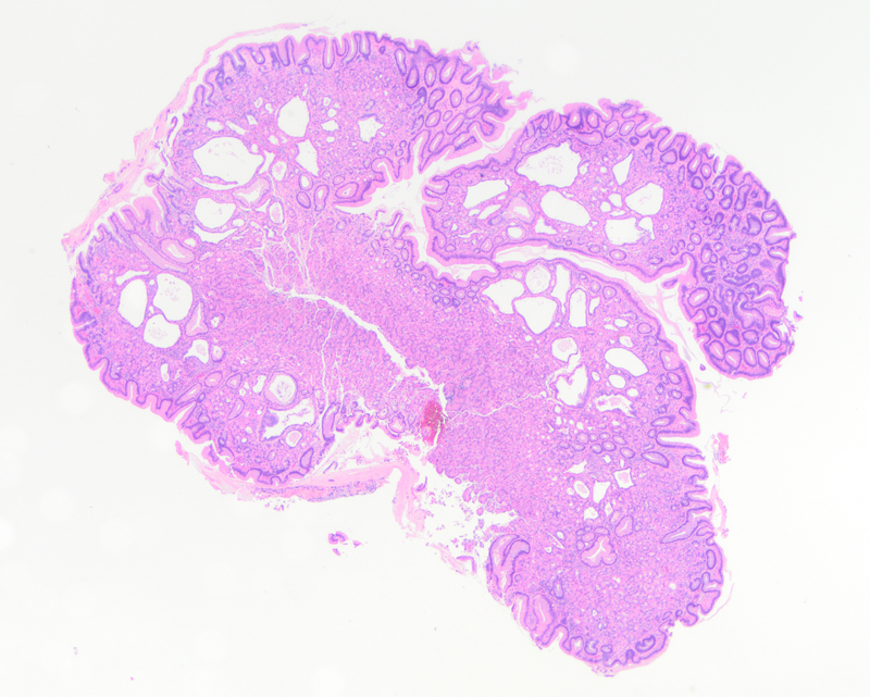
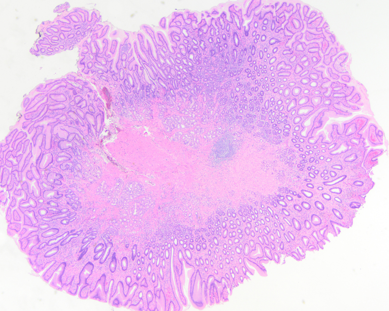
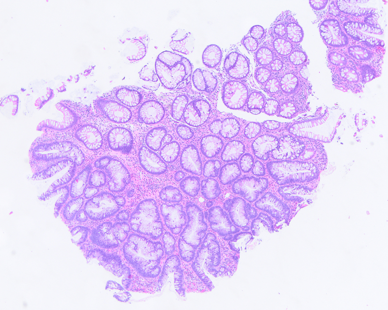
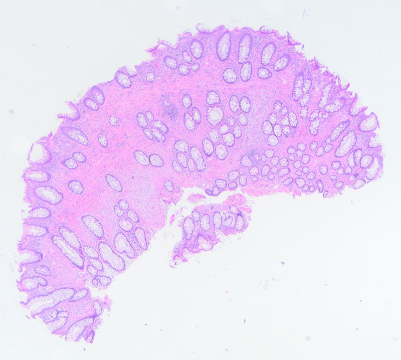
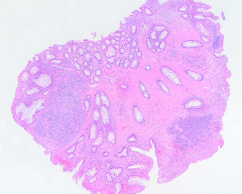
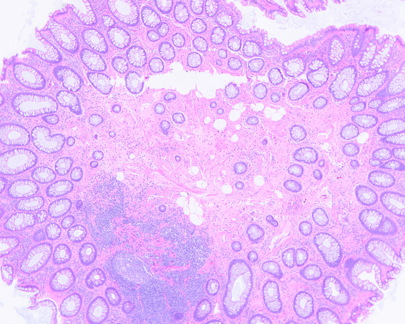
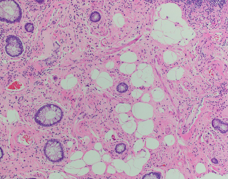
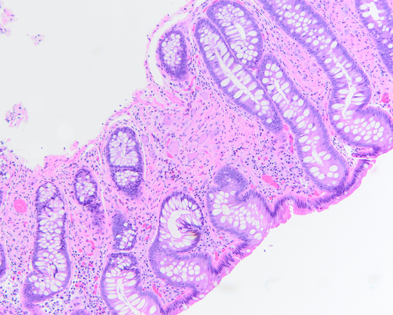
One polyp in the stomach consisted of elongated pits lined by foveolar epithelium (Figure 1) and a second polyp had cystically dilated glands lined by parietal cells and chief cells (Figure 2). Within the transverse colon, there was a polyp with crowded crypts composed of elongated, basally oriented nuclei (Figure 3). In the ascending and sigmoid colon there were several polyps characterized by benign glands separated by unusually prominent stroma within the lamina propria (Figures 4 and 5). The sigmoid colon was notable for a polypoid lesion composed of mature adipocytes at the basal aspect of the lamina propria (Figures 6 and 7). Lastly, additional colonic polyps had proliferations of ganglion cells and spindle cells between glands within the lamina propria (Figure 8).