Case of the Month: Painless Face Nodule
By Anna-Karoline Israel, MD (PGY-3), Glynis A. Scott, MD, and Dongwei Zhang, MD
Clinical history
An 82-year-old male presents with an enlarging, painless nodule in the right lateral malar cheek.
Past medical history
The patient has a history of basal cell carcinoma and a remote history of left-sided parotid gland lesion.
Recent history
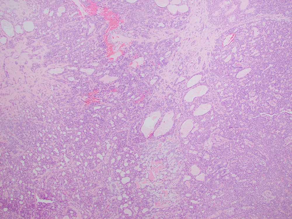
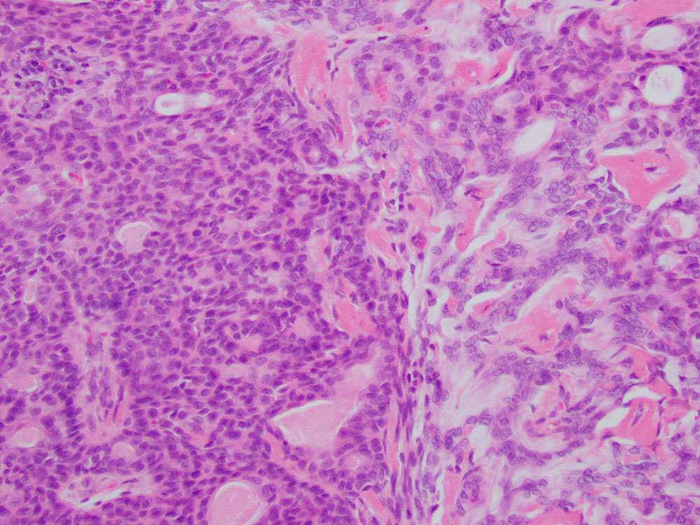
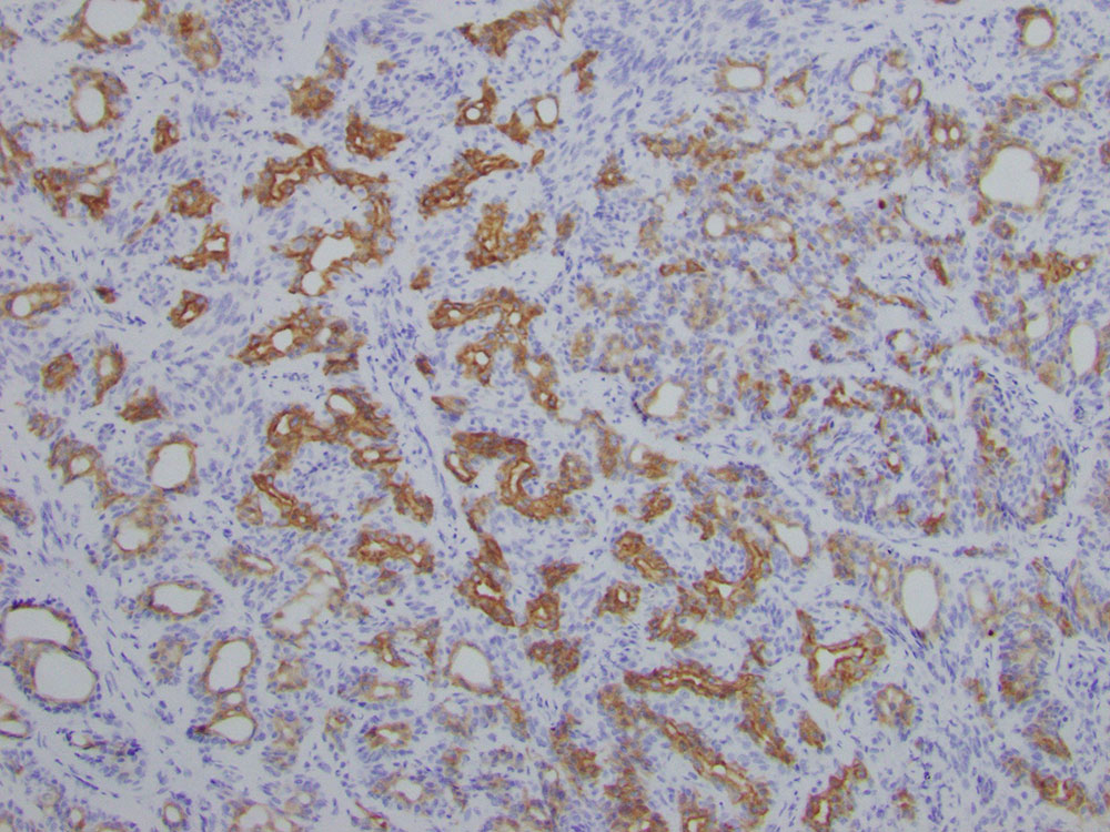
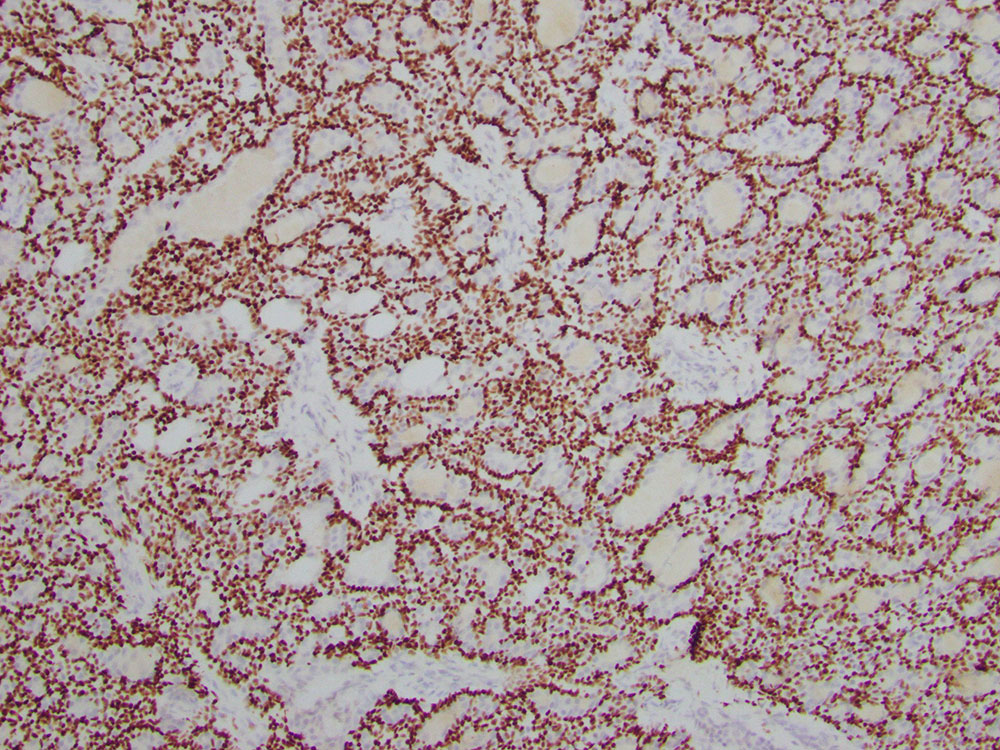
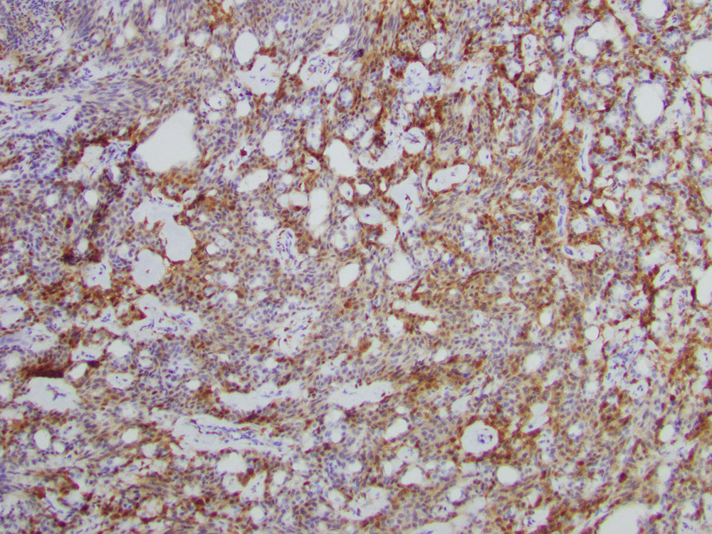
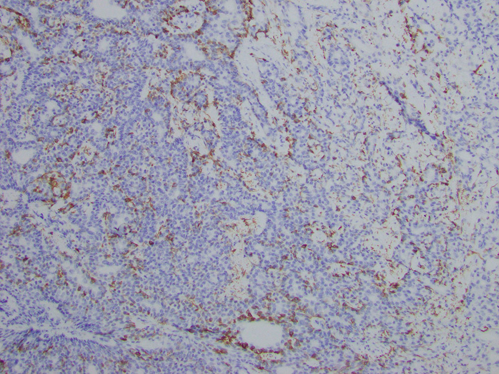
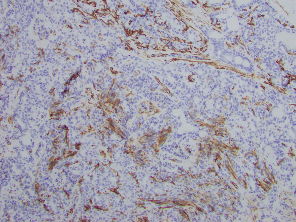
Sonographically, the lesion was located in the deep dermis and subcutaneous fat and appeared solid. Biopsy of the lesion was performed by a dermatologist. Histologic sections revealed a tumor with tubular and ductal structures lined by basaloid epithelial cells. Focally there was myxoid stroma present (Figures 1 and 2). Immunohistochemically, the neoplastic cells were positive for CAM 5.2 (Figure 3), p63, p40 (Figure 4), S-100 (Figure 5), and GFAP (focal, Figure 6), but negative for smooth muscle actin (SMA) (Figure 7). Rearrangement of the PLAG1 (8q12.1) locus was present in 96% of interphase cells by fluorescence in situ hybridization, supporting the diagnosis.