Case of the Month: Granular Esophageal Lesions
Authors: Anna Karoline Israel, MD, PGY-4, Diana Agostini-Vulaj, DO
Clinical History
A male in his 6th decade of life presented to the gastroenterology clinic for a repeat esophagogastroduodenoscopy (EGD) to assess for treatment response following his recent diagnosis of eosinophilic esophagitis managed with omeprazole and fluticasone.
Past Medical History
The patient has a history of asthma, chronic rhinitis, gastroesophageal reflux disease, biliary dyskinesia and hypercholesterolemia. For the past year, the patient noted abdominal pain, bloating, and heartburn; symptoms have improved since treatment was initiated.
Recent History
EGD findings included two granular areas in the proximal esophagus, 2-3 mm in size, which were biopsied. No furrows or rings were seen throughout the esophagus. Additional non-targeted biopsies from the upper and distal esophagus were obtained. The stomach showed small gastric polyps in the body. The remaining exam was otherwise unremarkable.
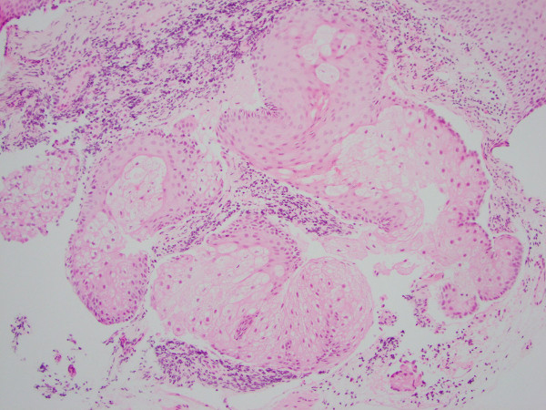
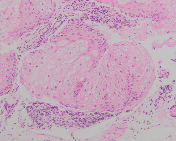
H&E sections of the non-targeted upper and distal esophageal biopsies were essentially unremarkable with no intraepithelial eosinophils seen. Sections of the granular areas in the proximal esophagus demonstrated lobules of large polygonal cells intimately admixed with squamous epithelium (Figs. 1-2) and surrounding chronic inflammation. On higher power, the cells were remarkable for distinct bubbly, vacuolated cytoplasm with a central variably scalloped nucleus, and no atypia (Fig. 3).