Case of the Month: Lung Nodule
Authors: Chauncey R. Syposs DO, MA; Andrew Evans MD, PhD; Moises Velez MD.
Clinical History
An elderly male presented to the hospital for workup of a left upper extremity soft tissue mass, concerning for sarcoma. Subsequent biopsy demonstrated an atypical B-cell proliferation. Excision of the soft tissue lesion demonstrated a nodular lymphoid proliferation with irregular centrocytes and large centroblasts, consistent with follicular lymphoma (W.H.O. Grade 2). Staging positron emission topography (PET) demonstrates a hypermetabolic lung nodule in the right lower lobe, which was biopsied. No other significant past medical history.
Recent History
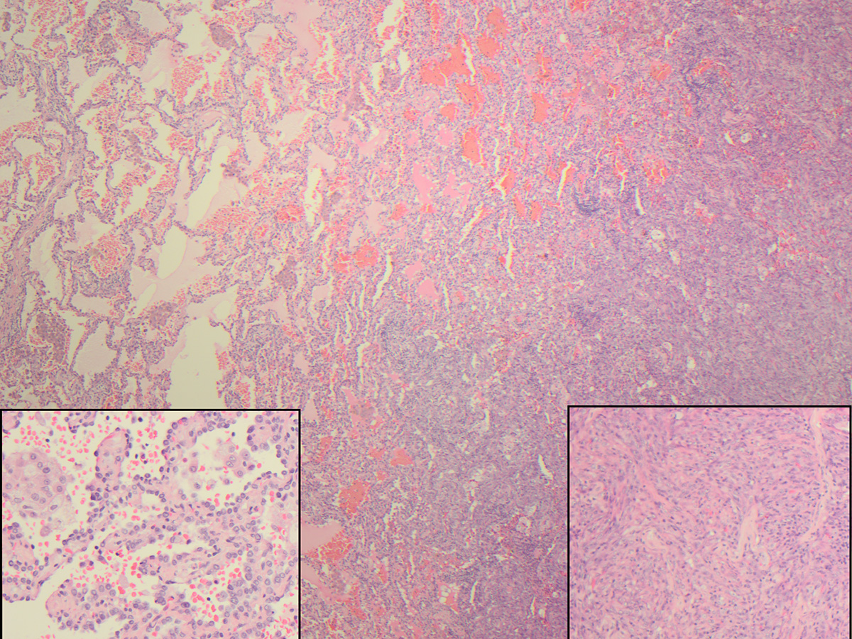
A CT-assisted lung biopsy was performed and was consistent with a diagnosis of histiocytic sarcoma, prompting a lobectomy of the right lower lobe with a wedge resection of the right middle lobe. H&E-stained slides demonstrate a biphasic tumor with sarcomatoid components and adenocarcinoma components (Figure 1). This tumor had two immunophenotypes: the adenocarcinoma component was positive for TTF-1, pan-cytokeratin, and napsin A, but negative for CD163; the sarcomatoid component was focally positive for pancytokeratin and CD163, but negative for TTF-1.