Equipment
Instruments Available for Structure Determination
-
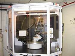
Bruker AXS X8 Prospector Ultra microfocus IµS sealed-tube X-ray generator featuring a highly sensitive charge coupled device (CCD) APEX II detector for rapid data acquisition and crystal analysis (MC 1-6811). The X-ray facility was recently upgraded in 2010 by an NIH/NCRR shared instrument grant (1S10 RR026501) worth $477,916 awarded to Dr. Joseph Wedekind.
-
The state-of-the-art system from Bruker AXS comprises an I(μ)S microfocus sealed tube Cu(Kα) X-ray source, integrated optics, and a highly sensitive APEX II CCD detector that can read out diffraction images in ˜1 sec. An Oxford 700-series cold stream is available to cool crystals to 100 K for data collection.

Close up of X8 Prospector I(μ)S microfocus sealed tube Cu(Kα) X-ray source, integrated optics, and APEX II CCD detector.
In addition, the upgraded system includes a point-and-click motorized goniometer head for rapid sample centering. The CCD detector is mounted on a 4-circle platform to rapidly obtain the most complete data sets. The 0.120 mm beam size produced by Bruker's multilayer Quazar optic is well suited for small crystals. The system can resolve a 295 Angstrom unit cell for challenging applications. A Mosquito liquid-handling robot (TTP LabTech) is available for rapid setup of nanoliter-scale crystallization experiments.
Nearby temperature-controlled rooms equipped with microscopes are available for crystal growth, documentation and mounting. The associated computational and stereographics facility houses several Linux and Mac workstations. Graphics and computational packages include: Coot, O, PyMol, CNS, CCP4, Phenix, SOLVE/RESOLVE, and HKL2000. The X-ray facility is located in the Department of Biochemistry & Biophysics. The executive director is Dr. Joseph Wedekind.
- Mosquito crystallization robot (TTP LabTech) for high-throughput, nanoliter-scale crystallization experiments (MC 1-6824).
- Microscopes for crystal viewing (MC 1-6807).
- Computers and software for structure determination and graphics (MC 1-6818).
- Dynamic Light Scattering (DLS) DynaPro Plate Reader II (Wyatt Technologies). Automated DynaPro Plate Reader II with onboard Domestic camera for industry standard microwell plates (96, 384, or 1536 well plates), fully integrated Dynamics software, and Peltier-driven temperature regulation that permits control of the plate from 4C - 85C with an ambient temperature of 24C. Dry gas required for operation below 20C. (MC 3-7418).
Instruments Available for Preparation of Vitrified Sample Grids for Cryo-EM
The Vitrobot™ Mark IV completely automates the vitrification process to allow for reproducible sample grid preparation which is a necessary for obtaining high quality images and 3D reconstructions. The software can precisely control the various steps along the vitrification process such as the blotting actions - application time, wait time, grid positioning
- A GloQube discharge system is available for the hydrophilization and cleaning of TEM grids before sample application and vitrification on the Vitrobot. The GloQube was purchased with funds from Professor’s Clara Kielkopf and Joseph Wedekind and the Department of Biochemistry & Biophysics
- Small sample volumes (~4 µL) can be applied by pipette tip through side ports available on both sides of the Vitrobot climate controlled chamber which is capable of maintaining 100% humidity. Grids then blotted and quickly plunged into a container of liquid Ethane surrounded by liquid nitrogen.
- Grids can be stored in liquid N2 and then transferred for screening and data collection on the university’s Electron Microscopy Core’s Thermo Fisher Talos L120C TEM or Hauptman-Woodward Institute’s Glacios (200 kV) cryo-TEM with a Volta phase plate, and Falcon 4 Detector
Instrument Available for Molecular Interaction Analysis
- BIAcore T200 for automated surface plasmon resonance (SPR) to attain high-quality kinetic or affinity data to quantify molecular binding interactions. Instrument sensitivity allows detection of macromolecular interactions, as well as macromolecular interactions with small molecules (>100 Da) (MC 3-8536). The BIAcore T200 purchase was funded by an NIH/NCRR shared instrument grant (1S10 RR027241) worth $386,596 awarded to Dr. Clara Kielkopf.
- VP-ITC (Microcal) isothermal titration calorimeter to determine the affinities and thermodynamic basis for macromolecular interactions.
- Fluoromax-3 fluorimeter to study macromolecular interactions using fluorescence.
Instrument Available for Determination of Molecular or Particle Size
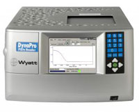
Wyatt technology’s DynaPro Plate Reader II instrument allows for high-throughput data collection of Dynamic Light Scattering (DLS, a.k.a. Quasi-elastic Light Scattering) data which can be used to determine the hydrodynamic radius (size) and size distribution of polymers and biopolymers in solution.
- Determine the hydrodynamic radius (Rh) and sample polydispersity (Survey multiple buffer conditions to find the buffer that gives the lowest %polydispersity)
- Determine the Critical Micelle Concentration (CMC) of surfactants under a variety of sample conditions (pH, temp, etc.)
- Plate reader accepts 96, 384, 1536-well glass bottom plates with approximate minimum sample volumes of 100, 50, and 5 µL respectively (Average read time for a full 384-well plate is <1 hour)
- Temperature controlled operation allows for the determination of polydispersity, size, and thermal stability between the range of 4- 85°C
- Onboard camera allows user to capture images of each well so images can be inspected for precipitant, bubbles, etc., which can lead to poor results
- The latest DYNAMICS software is available to collect, display, and analyze your data
The minimum concentration of the material under investigation depends on the molecular weight, the larger the polymer the lower the conc. that can be sampled. Using lysozyme (14 kDa) as an example a sample conc. as low as 0.125 mg/mL can be used whereas a protein with a molecular weight of ˜120 kDa could be used at a conc. of 0.015 mg/mL. We recommend starting with 0.5 - 1 mg/mL and trying multiple concentrations. The volume of sample needed depends on the plate used (see above) and the number of wells used (duplicate samples are recommended). Dr. Jenkins is available to help new users on the design, implementation, and analysis of DLS data on a fee-per-service basis.
Instrument Available for Purification of Proteins and Peptides
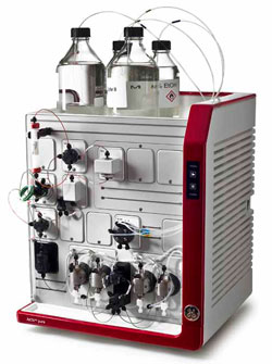
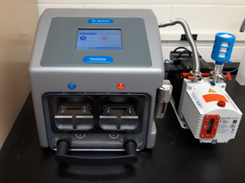
ÄKTA chromatography systems are robust user friendly systems widely used in both academic and industry labs. The ÄKTA Pure is a modular chromatography system for purification of proteins and peptides. The system is designed to allow for the easy addition of new modules as our purification needs evolve over time. The system supports a wide range of chromatography techniques and meets the automation requirements needed to deliver the highest purity. The ÄKTA’s user friendly UNICORN™ software has been designed to work with GE columns and media to meet any purification challenge.
- The Pure measures the UV absorbance at a fixed wavelength of 280 nm and is capable of a 0.001- 25 ml/min flow rate and has a max system pressure of 20 MPa
- The system accepts chromatography columns sold by GE and other companies such as Bio-Rad and maintains a library of commonly used columns to aid in purification method generation
- The ÄKTA has inlets for up to 4 buffers so it can equilibrate, elute, clean, and then prepare the column for storage without additional manual intervention
- Chromatography columns can be run in both directions to aid in cleaning and due to the unique column by-pass mode columns do not have to be removed to clean the system
- The system is equipped with a pH valve (can be taken off-line when not needed) that allows for simultaneous monitoring of pH and conductivity during such tasks as ion exchange chromatography
- The ÄKTA Pure and its fraction collector are stored in a 4 °C cold box and the fraction collector accepts 10-18 mm commonly used tubes such as Corning and Falcon 15 ml tubes
- The latest UNICORN software is available to collect, display, and analyze your data
- All knew ÄKTA users must undergo training with Dr. Jenkins on the instrument before being allowed to use the instrument alone, the amount of training depends on the users familiarity with ÄKTA systems
- Dr. Jenkins is available to help users with purification experiment design, implementation, and analysis of data on a fee-per-service basis
- The purchase of the ÄKTA Pure system was made possible through the support of the Center for AIDS Research (CFAR)
Instrument Available for Biomolecular Characterization
Circular dichroism (CD) is a useful technique for the characterization of biomolecules and the determination of absolute configuration and stereochemical analysis. JASCO Inc. J-1100 circular dichroism (CD) spectrometer (MC 3-8546) high scan speed allows one to measure samples quickly increasing productivity with the additional benefit is the minimal time exposure of biological samples to the high-energy UV light minimizing the risk of sample degradation. The dual polarizing prism optical design of the J-1100 results in stray light lower than 0.0003% enabling it to obtain high quality CD data even under conditions with high absorbance.
- Estimate secondary structure content. CD spectra have typical shapes for α-helical, β-sheet, or unfolded proteins as well as folded nucleic acids
- Non-destructively investigate the consequences of a protein or RNA mutation on its fold or structural stability
- Measure the thermal stability of a sample from a melting curve
- Temperature controlled operation via a single-position Peltier thermostatted cell holder for use with rectangular cells (Available temperature range -30 to +130 °C)
- Can accommodate cells of 10, 5, 2, 1 and sub-1 mm path lengths
- Cylindrical cells can be used but only in our non-Peltier cell holder
- For use with larger cuvettes an onboard built-in variable speed magnetic stirrer can be used to reduce precipitant, bubbles, etc., which can lead to poor results
- Perform chemical stability studies and investigate protein - protein interactions and ligand binding
- Light source is a air-cooled 150W Xenon lamp with a wavelength range: 180 – 600 nm
- The installed Spectra Manager II software suite is a comprehensive lab companion for capturing and processing data eliminating the need to learn multiple software programs
- The JASCO purchase was made possible through the support of the Departments of Chemistry and Biochemistry & Biophysics with a small equipment award from the URMC Scientific Advisory Committee
- The Facility has 3 shared use cuvettes of 0.1, 1, and 10 mm path length available for use for a fee (covers cleaning and replacement etc.) but we recommend users bring their own cuvette (~$170).
The CD requires purging with nitrogen gas BEFORE firing the lamp and this is critical to avoid damaging the optics. To avoid inadvertent use of the instrument before purging that would damage costly optics users should contact Dr. Jenkins before sign-up via the web-based calendar. This will insure he will be available to purge the instrument before their scheduled experiment.
The volume and concentration needed of material under investigation depends on the size of the cuvette and trying multiple concentrations is a good idea. For those working solely the far-UV CD region (190-250 nm) we recommend using smaller cuvettes (1 mm) and a good starting concentration would be ~0.2 mg/mL. Concentration is inversely proportional to the cuvette path length, so a 1mm path cuvette requires ~10x the concentration of a 10 mm cuvette. Higher sample concentrations are needed for near-UV CD (250-350 nm) spectra and possibly for estimating secondary structure content. Samples should contain as little chloride as possible, so NaCl and Tris buffers are not recommended. A good buffer for CD is usually low concentration (5-50 mM) phosphate or acetate with little or no salt (NaF can be tried as substitute for NaCl). Avoid using buffers with detergents, other molecules that contain aromatic groups, thiol reducing agents (DTT), and imidazole. Dr. Jenkins is available to help new users on the design, implementation, and analysis of CD data on a fee-per-service basis.
Instrument Available for Cell Lysis
The Avestin EmulsiFlex-C3 is a laboratory scale high pressure homogenizer commonly used in research laboratories and the pharmaceutical industry. It can be used with a small volume of lysate and is capable of obtaining both the low pressure necessary to rupture E. coli cells and the high pressures needed to lyse yeast cells. It can be used to make dispersions, emulsions, and produce liposomes but individual users will have to work out the experimental details specific to these applications but Dr. Jenkins can help with this on a fee-per-service basis.
- The EmullsiFlex-C3 has a usable operating pressure range of 2000 to 30,000 psi
- Multiple passes through the homogenizer at ~15000 psi will rupture E. coli. cells and more resistant cells including Pichia, and Saccharomyces can be lysed at higher pressures (~25000 psi)
- Cell suspensions must be free flowing and clump free before putting them in the instrument or it will clog and damage the instrument and result in loss of precious sample
- The minimum sample volume is 10mL (holdback volume of ~1mL) and the flow rate is ~50 mL/min and is independent of the homogenizing pressure selected
- To limit the risk of instrument damage it is recommend that the minimum sample volume be 20mL unless the user has significant experience
- Cold water and cold buffer can be used to cool sample flow path to offset the slow heat buildup that occurs during the homogenizing process
- Dr. Jenkins is available to train new users
- Users are expected to follow our instrument cleaning protocol and users are responsible for any damage to the equipment caused by negligent use