menu

News

20242023202220212020

Subscribe to Neuroscience LISTSERV

NGP Student Publishes First Author Paper in European Journal of Neuroscience

Tuesday, December 31, 2019

Emily WarnerEmily Warner, from the Padmanabhan labs, has authored a paper in European Journal of Neurosciences, "Sex differences in head?fixed voluntary running behavior in C57BL/6J mice".

Congratulations Emily!

Read More: NGP Student Publishes First Author Paper in European Journal of Neuroscience

URMC-099 Combats Surgery-Induced Delirium, Cognitive Dysfunction in Preclinical Model of Orthopedic Surgery

Wednesday, November 6, 2019

URMC-099

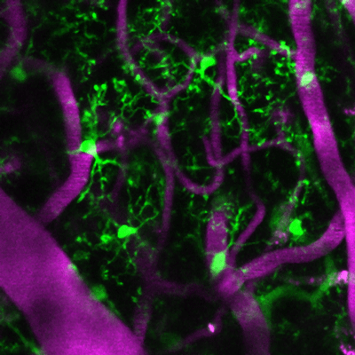
Living microglia, genetically marked to glow green, in the cerebral cortex with magenta colored blood vessels from a mouse treated with URMC-099.

A new study published in the Journal of Neuroinflammation found that prophylactic treatment with URMC-099 -- a "broad spectrum" mixed-lineage kinase 3 inhibitor -- prevents neuroinflammation-associated cognitive impairment in a mouse model of orthopedic surgery-induced perioperative neurocognitive disorders (PND).

PND, a new term that encompasses postoperative delirium, delayed neurocognitive recovery, and postoperative neurocognitive disorder, is the most common complication after routine surgical procedures, particularly in the elderly. Following surgery, such as hip replacement or fracture repair, up to 50 percent of patients experience cognitive disturbances like anxiety, irritability, hallucinations, or panic attacks, which can lead to more serious complications down the line. Currently, there are no FDA-approved therapies to treat it.

Developed in the laboratory of Harris A. "Handy" Gelbard, M.D., Ph.D., director of the Center for Neurotherapeutics Discovery at the University of Rochester Medical Center, URMC-099 inhibits damaging innate immune responses that lead to inflammation in the brain and accompanying cognitive problems. Using animal models of diseases like HIV-1-associated neurocognitive disorders, Alzheimer's disease and multiple sclerosis, Gelbard has shown that the compound blocks enzymes called kinases (such as mixed lineage kinase type 3, or MLK3) that respond to inflammatory stressors inside and outside cells.

Gelbard and Niccolò Terrando, Ph.D., director of the Neuroinflammation and Cognitive Outcomes laboratory in the Department of Anesthesiology at Duke University Medical Center, used an orthopedic surgery mouse model that recapitulates features of clinical procedures such as a fracture repair or hip replacement, which are often associated with PND in frail subjects. In a pilot experiment, they treated one group of these mice with URMC-099 before and after surgery, and another group prior to surgery only. Gelbard and Terrando's teams, including first author Patrick Miller-Rhodes, a senior pre-doctoral student in the Neuroscience Graduate Program working in the Gelbard lab at URMC, measured the following:

  • How the surgery affected the central nervous system and the immune cells (microglia) that reside there was evaluated using stereology and microscopy.
  • Surgery-induced memory impairment was assessed using the "What-Where-When" and Memory Load Object Discrimination tasks.
  • The acute peripheral immune response to surgery was assessed by cytokine/chemokine profiling and flow cytometry.
  • Long-term fracture healing was assessed in fracture callouses using micro-computerized tomography and histomorphometry analyses.
  • For additional details see the "Materials and Methods" section of the study

The team found that the surgery disrupted the blood brain barrier and activated microglia (a first line immune responder present in the inflamed brain), which led to impaired object place and identity discrimination when the mice were subject to the "What-Where-When" and Memory Load Object Discrimination tasks. Both URMC-099 dosing methods prevented the surgery-induced microgliosis (increase in the number of activated microglia) and cognitive impairment without affecting fracture healing.

"A major concern regarding the use of anti-inflammatory drugs for PND is whether they will affect fracture healing. We found that our preventive, time-limited treatment with URMC-099 didn't influence bone healing or long-term bone repair," said Gelbard and Terrando, professor of Neurology, Neuroscience, Microbiology and Immunology, and Pediatrics at URMC and associate professor of Anesthesiology at Duke University Medical Center, respectively. "These findings of improvement in cognition and normal fracture healing provide compelling evidence for the advancement of URMC-099 as a therapeutic option for PND."

"Right now we have nothing to treat this condition," said Mark A. Oldham, M.D., assistant professor in the department of Psychiatry at URMC who treats patients with PND. "We work hard to provide good medical care, including helping people sleep at night and making sure they are walking, eating and drinking, but it isn't clear that these efforts have any meaningful long-term impact."

According to Oldham, recent studies that track patients following an episode of PND show that many of them don't resolve completely, and that they have a new cognitive baseline after delirium.

"It is increasingly an accepted fact that after delirium, people have suffered some kind of neurological insult, which leaves them cognitively or functionally worse off than before the incident," he noted.

Next steps for the research include identifying definitive mechanisms for pain modulation, immune cell trafficking and neuro-immune characterization in PND. Gelbard and Terrando are tackling some of these questions with funds from the National Institutes of Health (RO1 AG057525). The current study was also funded by multiple grants from the NIH (P01MH64570, RO1 MH104147, RO1 AG057525 and F31 MH113504). The University of Rochester has four issued U.S. patents and multiple issued patents in foreign countries covering URMC-099.

Endocrine disruptors and male vulnerability to behavioral disorders

Tuesday, November 5, 2019

Marissa Sobolewski, Ph.D., says some chemicals can raise early-life testosterone levels in males, increasing risk of disorders such as ADHD.

Some neurobehavioral disorders — such as attention deficit hyperactivity disorder (ADHD), autism, and Tourette's syndrome — are far more common in males than females. But why?

During her talk at NIEHS Sept. 24, grantee Marissa Sobolewski, Ph.D., explained that such disparity exists partly because males experience a testosterone surge early in life.

She noted that endocrine disrupting chemicals (EDCs), which mimic sex hormones, can intensify that surge and change the epigenetic profiles of males' brains. Epigenetic changes, which are heritable, affect DNA function but not the underlying nucleotide sequence.

The big question Sobolewski seeks to answer is how disruption of early-life testosterone and brain epigenetics can increase males' risk of developing one of these disorders.

Read More: Endocrine disruptors and male vulnerability to behavioral disorders

NGP Student Publishes First-Author Paper in Journal of Neuroinflammation

Friday, November 1, 2019

Patrick Miller-Rhodes has had his paper, "The broad spectrum mixed-lineage kinase 3 inhibitor URMC-099 prevents acute microgliosis and cognitive decline in a mouse model of perioperative neurocognitive disorders" published in the Journal of Neuroinflammation.

Congratulations Patrick! Here is to many more.

Read More: NGP Student Publishes First-Author Paper in Journal of Neuroinflammation

Neuroscience Students and Faculty Receive Awards at Convocation

Monday, September 23, 2019

Neuroscience students and faculty made a big splash at the 2019 School of Medicine & Dentistry Convocation Ceremony. The following were awarded and recognized for their achievements:

  • Martha Gdowski, Ph.D., received the Manuel D. Goldman Prize for Excellence in First Year Teaching & the Marvin J. Hoffman Faculty Mentoring Award
  • John Olschowka, Ph.D., received a Commendation for First Year Teaching
  • Sergiy Nadtochiy, Ph.D., received a Commendation for First Year Teaching
  • Sarah McConnell, Ph.D., received the Gold Medal Award for Excellence in Teaching
  • Johanna Fritzinger, first year NGP student, received the Irving L. Spar Fellowship Award
  • Sarah Yablonski, first year NGP student, received the Merritt & Marjorie Cleveland Fellowship & the Robert L. and Mary L. Sproull University Fellowship
  • Suzanne Haber, Ph.D., received the Dean's Professorship
  • Marc Halterman, M.D., received the Dean's Associate Professorship

Congratulations to all recipients!

Convocation Awards 2019 - Neuroscience Dept.

You're Invited: URMC Reception at SfN

Wednesday, September 18, 2019

SfN Reception Invitation

Professor Maddox receives funding from NIH and NIDCD

Friday, August 23, 2019

Professor Ross Maddox has received funding from the National Institutes of Health (NIH) and the National Institute on Deafness and Other Communication Disorders (NIDCD) for his project, "Rapid acquisition of the frequency-specific auditory brainstem response through parallel stimulus presentation." The frequency-specific auditory brainstem response (ABR) is a diagnostic tool that is used to test for hearing loss in infants and prescribe treatments such as behavioral intervention or hearing aids. The ABR is performed while the baby sleeps, which leads to tests with unpredictable durations that are often too short to provide complete information. This project is relevant to public health because it provides a new kind of ABR exam that can be run much faster, allowing more complete and accurate diagnoses to be made.

NGP Student Article Published in the bioRxiv

Monday, August 19, 2019

Abstract: Perioperative neurocognitive disorders (PND), including delirium and postoperative cognitive dysfunction, are serious complications that afflict up to 50% of surgical patients and for which there are no disease-modifying therapeutic options. Here, we test whether prophylactic treatment with the broad spectrum mixed-lineage kinase 3 inhibitor URMC-099 prevents surgery-induced neuroinflammation and cognitive impairment in a translational model orthopedic surgery-induced PND. We used a combination of two-photon scanning laser microscopy and CLARITY with light-sheet microscopy to define surgery-induced changes in microglial morphology and dynamics. Orthopedic surgery induced microglial activation in the hippocampus and cortex that accompanied impairments in episodic memory. URMC-099 prophylaxis prevented these neuropathological sequelae without impacting bone fracture healing. Together, these findings provide compelling evidence for the advancement of URMC-099 as a therapeutic option for PND.

Read More: NGP Student Article Published in the bioRxiv

Translating Fear

Thursday, August 1, 2019

Fear is a normal emotion in animals and humans alike, enabling us to stay safe and avoid harm. Human anxiety disorders, including conditions such as post-traumatic stress disorder and social anxiety disorder, are conceptualized as an 'over-reactivity' of normal fear responses, or inability to modulate fear once the response is no longer appropriate. In the last decade, studies have delineated fear circuitry in rodents, to build models of how normal fear responses are instantiated when threat is present, and how fear learning is 'neutralized' by new learning when threat is no longer present (a process known as extinction).

Up until now, these models had not yet been examined in brains similar to those in humans. In a new study, published in Cerebral Cortex, researchers at the University of Rochester Medical Center used very small, paired injections of neuronal tracers in the brains of nonhuman primates, and examined how the amygdala communicates with the primate analogues of the prelimbic (fear) and infralimbic (extinction) PFC. The study, co-led by Keshov Sharma, a third year MSTP graduate student, found that, as in rodent, projections from the basal nucleus of the amygdala exist in nonhuman primates. However, there is also a substantial input from a neighboring region known as accessory basal nucleus, a greatly expanded amygdala nucleus in nonhuman primates and humans.

Read More: Translating Fear

Professor Carney receives renewed NIH funding

Monday, July 15, 2019

Congratulations to Professor Laurel Carney on the renewal of her NIH R01 for her project, "Auditory Processing of Complex Sounds." The Carney Lab hypothesizes that midbrain sensitivity to neural amplitude and frequency fluctuations in peripheral responses provides a robust representation of complex sounds, including speech. Aim 1 tests this hypothesis with physiological and behavioral studies of midbrain responses to stimuli that combine these cues, including "designer" stimuli with conflicting cues to determine how they may interact. These results will be used to test and refine a new computational model for midbrain responses with sensitivity to these cues. Aim 2 tests the hypothesis with physiological responses of midbrain neurons to voiced speech, to directly test model predictions based on characterization of each neuron's sensitivity to these cues. Understanding the role in speech coding of the amplitude and frequency fluctuations in peripheral responses is clinically significant because these fluctuations are vulnerable to SNHL. In Aim 3, we will test the hypothesis that amplitude and frequency fluctuations can be manipulated in synthetic speech to influence intelligibility in human listeners with or without SNHL, in quiet and in noise. Preliminary results from modeling, physiological, and behavioral studies support the proposed hypotheses. Ultimately, our goal is to extend this approach to manipulate fluctuation contrasts in running speech, to effectively "correct" sound for the impaired ear.

Brain stimulation speeds up visual learning in healthy adults, helps patients re-learn how to see

Tuesday, May 28, 2019

Practice results in better learning. Consider learning a musical instrument, for example: the more one practices, the better one will be able to learn to play. The same holds true for cognition and visual perception: with practice, a person can learn to see better—and this is the case for both healthy adults and patients who experience vision loss because of a traumatic brain injury or stroke.

The problem with learning, however, is that it often takes a lot of training. Finding the time can be especially difficult for patients with brain injuries who may, for instance, need to re-train their brains to learn to process visual cues.

But what if this learning process could be accelerated?

That's what University of Rochester researchers Duje Tadin, a professor of brain and cognitive sciences, and Krystel Huxlin, the James V. Aquavella, M.D. Professor in Ophthalmology at the University's Flaum Eye Institute, set out to determine. Motivated by emerging evidence that brain stimulation might aid learning, Tadin and Huxlin collaborated with researchers at the Italian Institute of Technology to study how different types of non-invasive brain stimulation affect visual perceptual learning and retention in both healthy individuals and those with brain damage. Their results, published in a paper in the Journal of Neuroscience, could lead to enhanced learning efficacy for both populations and improved vision recovery for cortically blind patients.

Enhancing learning with brain stimulation

Learning is difficult and often takes a long time, Tadin says, "because after early childhood our brains become less plastic." The brain's ability to change and reorganize itself decreases as a person ages, so learning new tasks, or re-learning tasks after experiencing a brain injury, becomes more challenging.

To test if and how visual perceptual learning might be accelerated, researchers presented study participants with a computer-based task. Participants were shown clouds of dots and were asked to determine which way the dots moved across the computer screen. The task measured the participants' motion integration threshold; motion perception is important in enabling people to see movement and either to avoid or interact with moving objects. Participants were then asked to perform the task while sub-groups were given different types of brain stimulation, each involving a non-invasive electrical current applied over the visual cortex. The researchers found that one particular type of brain stimulation, called transcranial random noise stimulation (tRNS), had remarkable effects on improving participants' motion integration thresholds when they performed the task.

"All groups of participants got better at the dot motion task with practice, but the group that also trained with tRNS improved twice as much and was able to learn the motion task better than other groups," Tadin says.

Surprisingly, the researchers also found that when they re-tested the participants six months later, the boosts in performance were still there: the participants in the tRNS group had retained what they had learned and were still able to do better on the motion task compared to the groups that were given other stimulation techniques or training alone.

Read More: Brain stimulation speeds up visual learning in healthy adults, helps patients re-learn how to see

In The News: Interviews with Scientists: Rianne Stowell

Tuesday, May 7, 2019

Rianne StowellRianne Stowell, fifth year graduate student in the Majewska Lab, is interviewed by Hello Bio about her research, the most important lesson she's learned as Ph.D. student, her proudest career moment and more.

Read More: In The News: Interviews with Scientists: Rianne Stowell

Dr. Gary Paige is awarded 2019 Peter Shrager Award

Friday, May 3, 2019

Gary PaigeDr. Gary Paige is the 2019 recipient of the Peter Shrager Award, which recognizes exceptional service to the Rochester neuroscience community. The award was established in 2017 to honor Dr. Peter Shrager, professor in the Department of Neuroscience, for his commitment to the education and research mission of the neuroscience enterprise at the University over his last 40 years as a faculty member. Dr. Gary Paige served as the chair of Neurobiology and Anatomy for more than ten years and was a tireless advocate for neuroscience faculty and students. In this role, he built a very open neuroscience community, expanding the department through secondary appointments and encouraging faculty be active members of the neuroscience community at large. He was a strong supporter of graduate and medical education, using creative solutions to forge collaborative and interdisciplinary research that integrated basic, translational and clinical approaches. Since stepping down as chair, he has continued to be influential in creative ways by marrying his two loves—art and neuroscience—in the Arts in Mind project, an educational experience for UR students and faculty. Dr. John Foxe presented the award at the annual neuroscience retreat on Friday, April 12.

NGP Alum, Aleta Steevens, awarded Doty prize

Friday, May 3, 2019

Aleta Steevens

Aleta Steevens, recent doctoral graduate from the Kiernan lab, received the Robert Doty prize for the 2019 outstanding dissertation in neuroscience. The Doty prize is named in the honor of longtime faculty member Robert Doty, who made great contributions to neuroscience research at the University of Rochester and nationally. It is awarded on the basis of the impact and importance of research, novelty of experimental design, independence and creativity of the student and research implications and relevance for neuroscience. Aleta's thesis entitled "The Dynamic Role of SOX2 in Mammalian Inner Ear Development," which she successfully defended on April 16, 2018, embodied all these characteristics. Aleta was also awarded an NIH predoctoral fellowship, and her work has resulted in two first-author publications. Beyond her research, Aleta was exceptionally active and successful in teaching. She served as a TA in the course Biology of Mental Disorders, and won the Edward Peck Curtis teaching award in 2016. Aleta has now moved to her postdoctoral position with Dr. Walter Low at the University of Minnesota. Dr. Peter Shrager presented the prize at the annual neuroscience retreat on Friday, April 12.

Handy Gelbard Honored for Pediatric HIV/AIDS Research

Wednesday, April 17, 2019

Harris Gelbard

Handy Gelbard, M.D., Ph.D., professor and director of the Center for Neurotherapeutics Discovery at URMC, is the 2019-2020 recipient of the Herman and Gertrude Silver Award, which honors individuals who have made significant contributions in the field of pediatric HIV and AIDS. The award is given by the Children's Hospital of Philadelphia (CHOP) and the Department of Pediatrics of the Perelman School of Medicine at the University of Pennsylvania. Past award winners include a Nobel laureate and HIV investigators from leading academic institutions, the National Institutes of Health (including the current directors of the Office of AIDS Research and the National Institute of Allergy and Infectious Diseases), and the Centers for Disease Control and Prevention.

For the past 10 years Gelbard's lab has been developing a compound called URMC-099, which dampens inflammation and has shown promise in reversing the neurological problems associated with HIV. Children with HIV who are taking combination antiretroviral therapies are extremely vulnerable to inflammation; the developing nervous system is of particular concern, as inflammation in the brain can lead to major cognitive problems.

The possibility of a new class of therapies that reduces the burden of neuroinflammation and supports normal synaptic architecture (the basis for learning and memory) offers considerable hope for children that are saddled with the unwanted burden of HIV, despite effective control of the virus.

Gelbard believes the path forward for URMC-099 as an adjunct agent for children living with HIV and neurologic disease will likely involve combination therapy with next generation antiretroviral agents. This is a priority in resource-limited settings such as Africa, and Gelbard is working with David Bearden, M.D., assistant professor in the division of Child Neurology at URMC to help advance uses for URMC-099 in pediatric patients there. Bearden's work is supported by a National Institute of Neurological Disorders and Stroke grant to Gretchen Birbeck, M.D., M.P.H., professor of Neurology and Michael Potchen, M.D., professor of Imaging Sciences. The work is also supported by the University of Rochester Center for AIDS Research.

Gelbard will receive the Silver Award in November during a two-day symposium at CHOP. He will present pediatric grand rounds describing his progress in inventing the class of compounds spearheaded by URMC-099 and its role in treating pediatric and adult HIV infection and its complications. He'll also give a seminar on current and future developments related to URMC-099 to attendees from multiple medical and scientific institutions in Philadelphia.

Brendan Whitelaw receives 2019 Edward Peck Curtis Awards for Excellence in Teaching by a Graduate Student

Tuesday, April 16, 2019

Brendan Whitelaw Curtis AwardNeuroscience Graduate Program and Medical Scientist Training Program student Brendan Whitelaw has been selected as one of the recipients of the 2019 Edward Peck Curtis Awards for Excellence in Teaching by a Graduate Student. The awards were established to recognize graduate students who advance the teaching mission of the University by providing highly skilled and innovative undergraduate education. Students were nominated by their department chair and a faculty member. Congratulations, Brendan!

Read More: Brendan Whitelaw receives 2019 Edward Peck Curtis Awards for Excellence in Teaching by a Graduate Student

An eye for seeing big problems, and solving them

Friday, April 12, 2019

From the very beginning of her career, Krystel Huxlin has looked for big problems to solve. Now she's doing something about a very big problem indeed: Loss of vision after a stroke or brain injury.

"When people have a stroke that affects the motor system, they are sent to rehab as soon as possible," says Huxlin, the James V. Aquavella, M.D. Professor in Ophthalmology at the University of Rochester's Flaum Eye Institute. "They get retrained to use a weak arm or leg, and to regain speech."

"But if they have a stroke that affects vision, which involves more than 30 percent of our brain matter and is our dominant sense, they are told there's essentially nothing that can be done." They're told "they should go home and get used to being blind."

Even more frustrating for Huxlin is when applications for disability are challenged because a stroke or injury victim "still has" a 50 percent field of vision. With only a 50 percent field of vision, it's difficult to spot a car coming from the right or left, to detect objects, to navigate through an airport, and even--especially if the "blind area" is to the right of center—to keep your eye anchored on the line of a book you are trying to read.

"All of these things become excruciatingly difficult," Huxlin says.

Read More: An eye for seeing big problems, and solving them

University of Rochester Investigators Attend the 2019 ARO MidWinter Meeting

Friday, February 22, 2019

The University of Rochester was well-represented at the Association for Research in Otolaryngology (ARO)'s 42nd Annual MidWinter Meeting. This prestigious academic conference brings together researchers and clinicians from around the world to present their work on diverse topics in the fields of Otolaryngology and Auditory Neuroscience. Several UR Neuroscience Department investigators were on hand to share their findings with over 1500 colleagues at Baltimore's Marriott Waterfront from February 9th to the 13th.

Genetic signaling pathways underlying cochlear development, damage, and recovery were highlighted by the White and Kiernan labs. Patricia White's lab presented their work on hearing recovery within adult mouse cochlea after noise damage. Jingyuan Zhang, a postdoctoral candidate, proposed activation of the ErbB signaling pathway as a means to promote hearing recovery and regeneration in the inner ear. Holly Beaulac, a Neuroscience graduate student, discussed how the transcription factor FOXO3 may play a role in cochlear inflammatory regulation.

Felicia Gilels, a CBD graduate student in Amy Kiernan's lab, examined the role of the Notch signaling pathway in cochlear maturation. Specifically, she presented work showing that loss of JAG1 leads to auditory neuropathy, caused by stereocilia defects in the inner hair cells. These results indicate that hair cell regeneration by manipulating Notch should proceed cautiously, as clearly the postnatal roles of Notch are not fully understood.

Ross Maddox's lab investigates how multiple sensory stimuli can be processed and how distractions including noise can affect the efficacy of such input. Lab members Madeline Cappelloni, a BME graduate student, and undergraduate Sara Fiscella explained the ways in which coherence of visual and auditory stimuli may be disrupted during task manipulation.

See Ross Maddox's research elevator pitch here: Ross Maddox - #pitchARO

Yingxuan Wang, a BME graduate student in Ken Henry's lab, described their investigation into inferior colliculus neural responses when birds attempt to detect tones in noise. Laurel Carney and her students showcased their findings across five posters. BME graduate student Langchen (Elsie) Fan provided physiological model simulations of comodulation masking release, or the ways tones can become easier to hear in noise, for individuals with and without hearing loss. Paul Mitchell, a BME graduate student, helped characterize midbrain selectivity for direction and velocity during sound presentation.

In the realm of vestibular research, J. Chris Holt gave a podium talk on establishing the pharmacological tools to investigate the physiological role of mammalian vestibular efferents, the nerves that project from the central nervous system to the vestibular sensory cells. Benjamin Crane and undergraduate Raul Rodriquez discussed the adaptation of heading perception which is important for ambulation, navigation, and spatial orientation. Daniel Martin, a medical resident in the Crane lab, presented his use of a novel virtual reality technique to adapt heading perception in cases of unilateral vestibular loss.

Art and science intersected in the neuroscience of music presentations. Using EEG, Nathaniel Zuk, a postdoctoral candidate in Edmund Lalor's lab, revealed specialized high-level processing of both speech and music. Anne Luebke featured three posters focused on how musical training can influence one's ability to discriminate between tones in noise. She also highlighted ongoing collaborative work with Loisa Bennetto and Paul Allen on how musical aptitude can predict auditory filtering in young adults and children with Autism Spectrum Disorder.

ARO's 2019 MidWinter Meeting provided an impressive showcase of promising areas of research in otolaryngology and auditory neuroscience. We expect UR's scientific impact to continue at next year's meeting in San Jose, California!

PHOTOS:

ARO's Mid-winter meeting banner

ARO's Mid-Winter Annual Meeting

Jingyuan Zhang, PhD at her Podium Talk with Patricia White, PhD moderating

Jingyuan Zhang, PhD at her Podium Talk with Patricia White, PhD moderating

Ken Henry, PhD and Yingxuan Wang, PhD Candidate - BME, at their poster

Ken Henry, PhD and Yingxuan Wang, PhD Candidate - BME, at their poster

Langchen (Elsie) Fan, PhD Candidate - BME, at her poster

Langchen (Elsie) Fan, PhD Candidate - BME, at her poster

Paul Mitchell, PhD Candidate - BME, at his poster

Paul Mitchell, PhD Candidate - BME, at his poster

J. Chris Holt, PhD at his Podium Talk

J. Chris Holt, PhD at his Podium Talk

Jenna Glatzer (BS - Neuroscience ’18), Aleta Steevens (PhD - Neuroscience ’18), Courtney Kellogg (PhD Candidate - CBD), Felicia Gilels (PhD Candidate - CBD) Jingyuan Zhang (PhD - Biology ’18), & Holly Beaulac (PhD Candidate- Neuroscience)

Jenna Glatzer (BS - Neuroscience '18), Aleta Steevens (PhD - Neuroscience '18), Courtney Kellogg (PhD Candidate - CBD), Felicia Gilels (PhD Candidate - CBD) Jingyuan Zhang (PhD - Biology '18), & Holly Beaulac (PhD Candidate- Neuroscience)

Multi-Target Drugs Should Be in the Pharma Pipeline along with Precision Drugs

Thursday, January 31, 2019

"Precision medicine—providing the right treatment, for the right patient, at the right time—is saving lives. The use of therapies that home in on single targets is helping beat tough-to-treat diseases that were often deadly in the past," writes Harris Gelbard, a professor of neurology, pediatrics, neuroscience, and microbiology and immunology, who is also director of the Medical Center's Center for Neurotherapeutics Discovery. "But we're overlooking another class of extremely important and promising candidates: multi-target drugs."

Read More: Multi-Target Drugs Should Be in the Pharma Pipeline along with Precision Drugs

Study suggests how high blood pressure might contribute to Alzheimer’s

Monday, January 28, 2019

The brain's system for removing waste is driven primarily by the pulsations of adjoining arteries, University of Rochester neuroscientists and mechanical engineers report in a new study. They also show that changes in the pulsations caused by high blood pressure slow the removal of waste, reducing its efficiency.

This might explain the association between high blood pressure and Alzheimer' disease, the researchers say. Alzheimer's, the most common cause of dementia among older adults, is characterized by abnormal clumps and tangled bundles of fibers in the brain.

The study, reported in Nature Communications, builds upon groundbreaking discoveries about the brain's waste removal system by Maiken Nedergaard, co-director of the University's Center for Translational Neuromedicine. Nedergaard and her colleagues were the first to describe how cerebrospinal fluid is pumped into brain tissue and flushes away waste. Subsequent research by her team has shown that this glymphatic waste removal system is more active while we sleep and can be damaged by stroke and trauma.

This latest research shows "in much greater depth and much greater precision than before" how the glymphatic system functions in the perivascular spaces that surround arteries in the outer brain membrane, says Douglas Kelley, an assistant professor of mechanical engineering and an expert in fluid dynamics. His lab is collaborating with Nedergaard's team as part of a $3.2 million National Institute on Aging grant.

For this study, Humberto Mestre, a PhD student in Nedergaard's lab, injected minute particles in the cerebrospinal fluid of mice, and then used two-photon microscopy to create videos showing the particles as they moved through the perivascular spaces.

Read More: Study suggests how high blood pressure might contribute to Alzheimer’s

In The News: UR study on brain waves may allow doctors to diagnose autism earlier

Friday, January 25, 2019

The following is an except of an article by Josh Navarro that originally appeared on WROC/RochesterFirst.com:

A new study to help understand brain waves in children with autism is underway right now at the University of Rochester Medical Center. Their aim is to foster earlier detection and foster better therapies in the future.

Children with autism respond differently when they hear a sound such as music or see an illustration. Honing in the difference in brain waves between autistic children and children who do not have autism, is part of a new study at Del Monte Institute for Neuroscience at URMC.

"If you can provide a biological marker that could be reproduced earlier in a child's development, then that therapy can start earlier, the better outlook for that particular child," said Dr. Evan Myers, Postdoctoral Fellow at the Cognitive Neurophysiology Lab in the University of Rochester Del Monte Institute for Neuroscience.

Researchers will place a electroencephalography cap and have kids observe different images on a computer screen. The findings will determine the next step through clinical trials with the goal of diagnosing a child with autism a lot sooner.

Read More: In The News: UR study on brain waves may allow doctors to diagnose autism earlier

Dr. Kuan Hong Wang comes to the University of Rochester

Monday, January 21, 2019

We are pleased to welcome Dr. Wang to the University of Rochester Medical Center, the Department of Neuroscience and the Del Monte Institute for Neuroscience from the NIH.

Dr. Wang comes to us as the former chief of the Unit on Neural Circuits and Adaptive Behaviors at the National Institute of Mental Health. Dr. Wang received his B.A. in Biochemical Sciences from Harvard College and his Ph.D. from the University of California at San Francisco, where he studied the molecular regulators of sensory axon growth and branching during development with Marc Tessier-Lavigne. Dr. Wang obtained postdoctoral training with Susumu Tonegawa at the Massachusetts Institute of Technology, where he examined the ways in which cortical neurons respond to an animal's experience by directly visualizing the molecular activity of a given set of neurons over several days in the live animal. With this approach, he revealed a physiological function of neural activity regulated gene Arc in sharpening stimulus-specific responses in visual cortex.