Non-Invasive Measures of Bone Health and Quality
Osteoporosis is a global epidemic with a socioeconomic burden that escalates progressively with the increase in life expectancy of the aging population. Women over the age of 40 are twice as likely to be osteoporotic than men of comparable ages. The risk of this disease nearly doubles every decade of life, such that nearly a quarter of women over 65 years of age are osteoporotic. The clinical consequences of this multifactorial disease, which is characterized by bone loss and low bone mineral density, include fragility fractures that lead to life-altering morbidity and premature mortality.
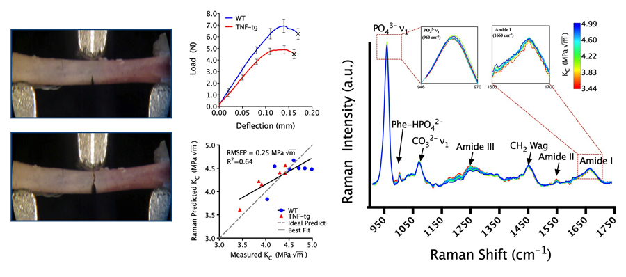
Bone fragility and fracture risk are conventionally assessed by measuring the bone mineral density (BMD) using dual-energy X-ray absorptiometry (DXA). However, while BMD correlates with bone strength and rigidity in animals and humans, it is nevertheless a poor predictor of fragility fracture risk, in part because the latter is poorly defined. Alternatively, fracture toughness can be a defined quality of bone since it assesses the bone’s resistance to crack propagation and subsequent fracture. Our hypothesis is that we could reliably predict fracture toughness by augmenting measurements from DXA and µCT with Raman spectroscopy.
As an alternative to standard medical imaging, Raman spectroscopy is a non-destructive vibrational spectroscopy technique that can reveal important compositional information about bone matrix and is sensitive to chemical and structural changes in the matrix. Raman spectroscopy has been used to study the chemical composition of bones, the effect of glucocorticoid in rheumatoid arthritis, aging bone and healing, among many other applications in bone but has yet to be used clinically. Some specific foci of research include:
- Adapting and validating spatially offset Raman spectroscopy (SORS) to measure the chemical composition of bone transcutaneously, using a mouse model of inflammatory-erosive arthritis and osteoporosis (TNF-alpha transgenic mice treated with systemic glucocorticoids).
- Multimodal assessment of bone quality to reliably predict ovariectomy-associated changes (to simulate menopause) in fracture toughness.
- Machine learning algorithms to diagnose osteoporosis and classify patients at risk of fragility fracture based on planar radiographs, DXA, and Raman spectroscopy.
Collaborators: Andrew Berger, Constantinos Ketonis, Warren Hammert
Funding: NIH/NIAMS R01 AR070613