Pathology Case of the Month: Pancreatic Mass
By Raman Baldzizhar, PGY-3 Resident
Clinical History
The patient is a 61-year-old male with vague upper abdominal discomfort and early satiety during the last few months. His past medical history is significant for hyperlipidemia, chronic back pain, GERD, and BPH. His comorbidities are under good control on medications and he has unremarkable past surgical history.
Imaging
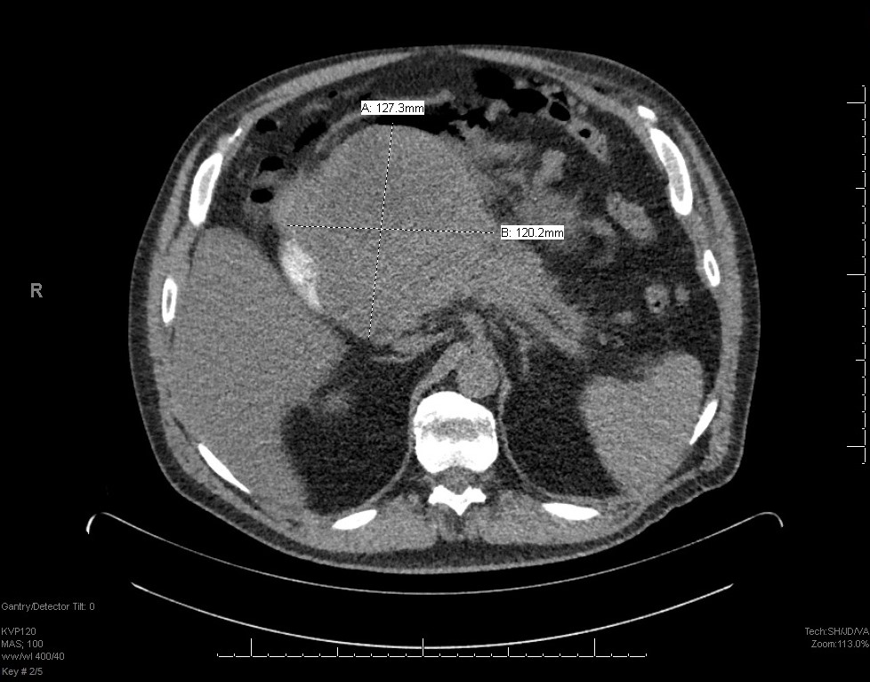
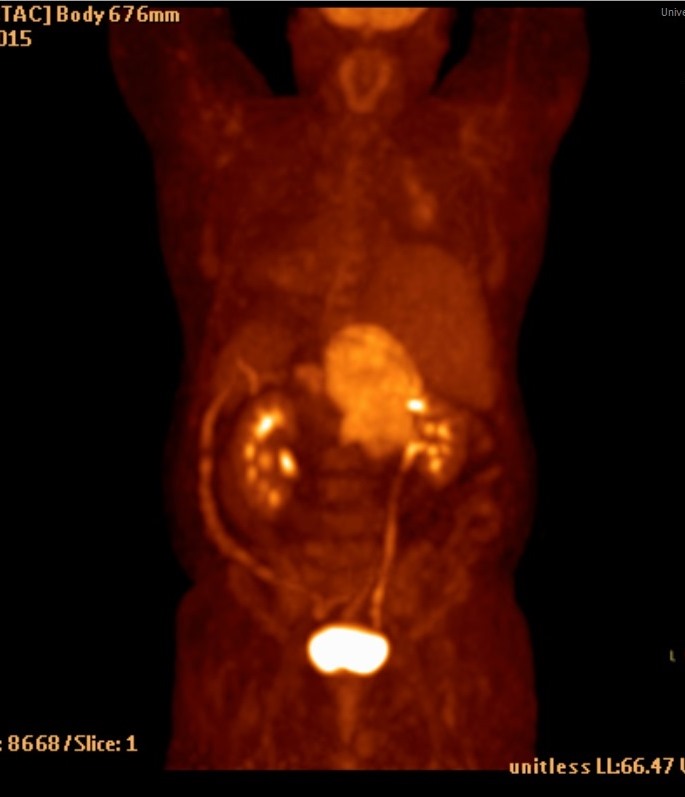
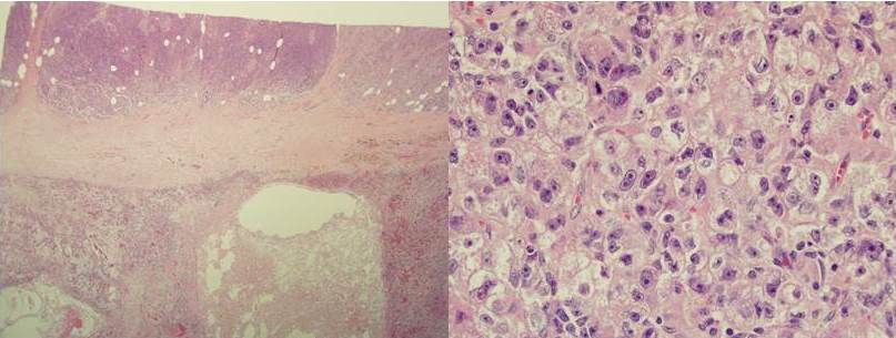
CT demonstrated an 18.6 cm complex mass in the mid-abdomen possibly arising from pancreas or bowel (Fig. 1). No significant adenopathy is identified. PET/CT demonstrated a large hypermetabolic mass centered at head of pancreas with involvement of the pancreatic body (Fig. 2). Following neoadjuvant treatment (six cycles of FOLFIRINOX chemotherapy) the patient underwent a pancreatoduodenectomy with right hemicolectomy and partial omentectomy. Sections from the pancreatic mass are shown in Figure 3.
Microscopic Examination
Sections from the pancreatic mass show sheets of cells without lumen formation, no desmoplastic stromal response. Uniform nuclei with vesicular chromatin and single prominent central nucleolus. Moderate finely granular, eosinophilic to amphophilic cytoplasm.