Case of the Month: Worsening Right Ankle Pain
Case Authors: Dingani Nkosi, MBBS, PhD (PGY-2), Aaron R. Huber, DO
Clinical History
A middle-aged man with 5 months of worsening pain above his right ankle was referred for orthopedic surgery evaluation. His pain was throbbing in nature, rated 6/10, did not radiate, and was exacerbated with activity and alleviated with rest. He had started using a cane when ambulating significant distances and also noticed a limp in his gait secondary to pain.
Past Medical History
He had no reported history of any injuries.
Recent History
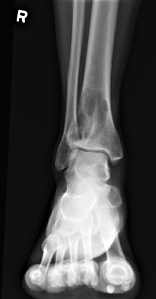
Radiographic film of the right ankle showed 5 cm eccentric lytic lesion involving the right distal tibia with features of cortical destruction and endosteal scalloping extending close to the articular surface (Figure 1). Nuclear medicine bone scan showed focal uptake in the known lytic lesion in the distal right tibia. The patient had an excisional biopsy to establish the diagnosis and subsequently had the mass excised with bone curettage.
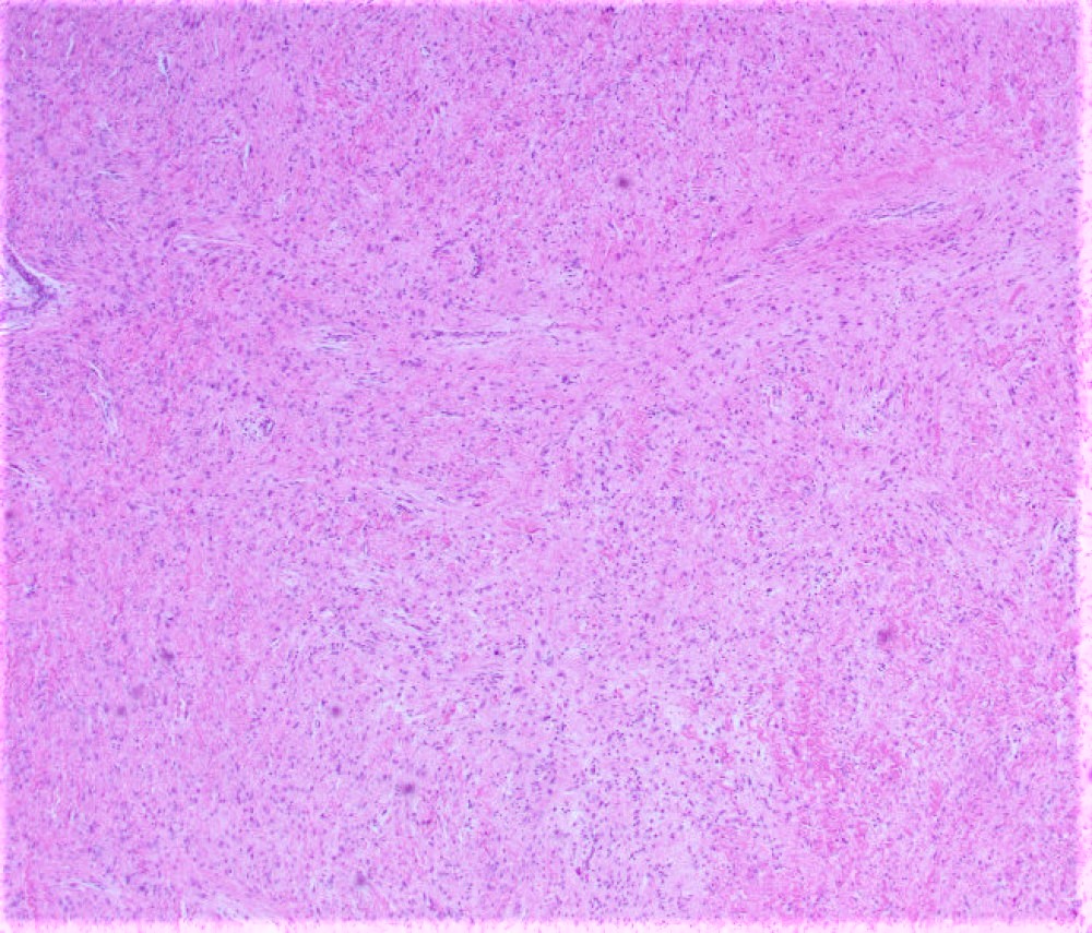
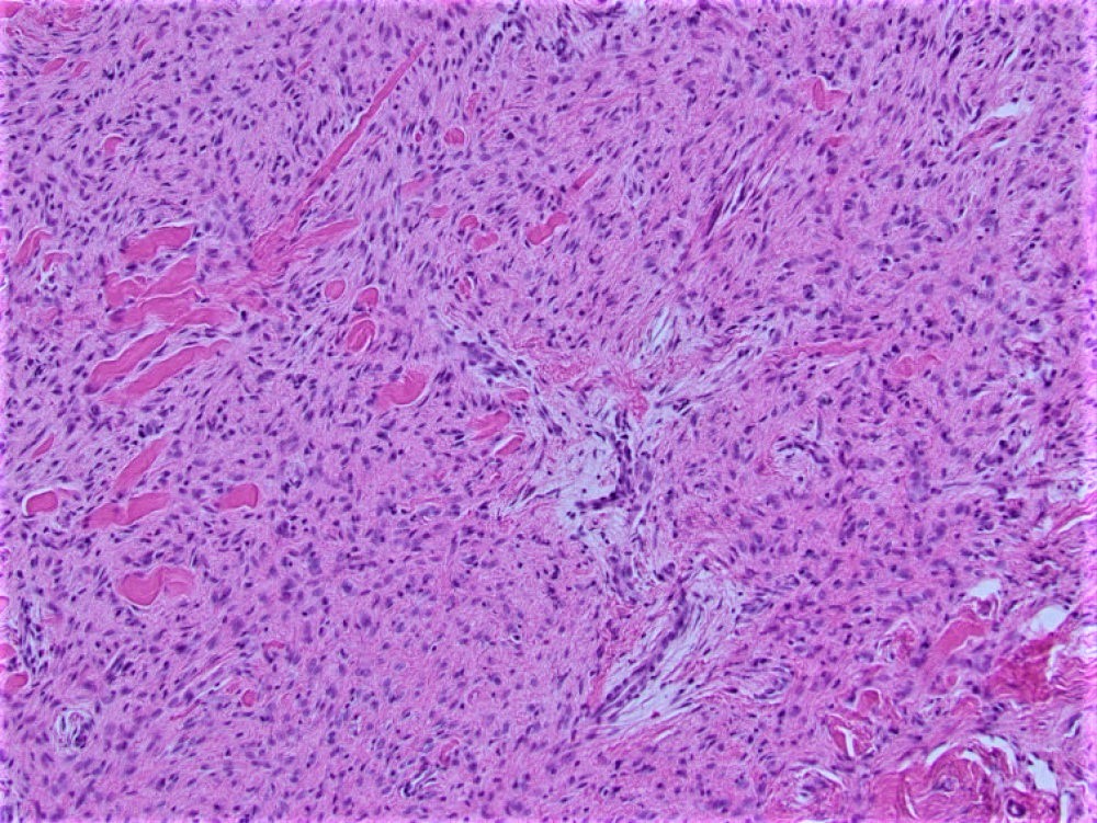
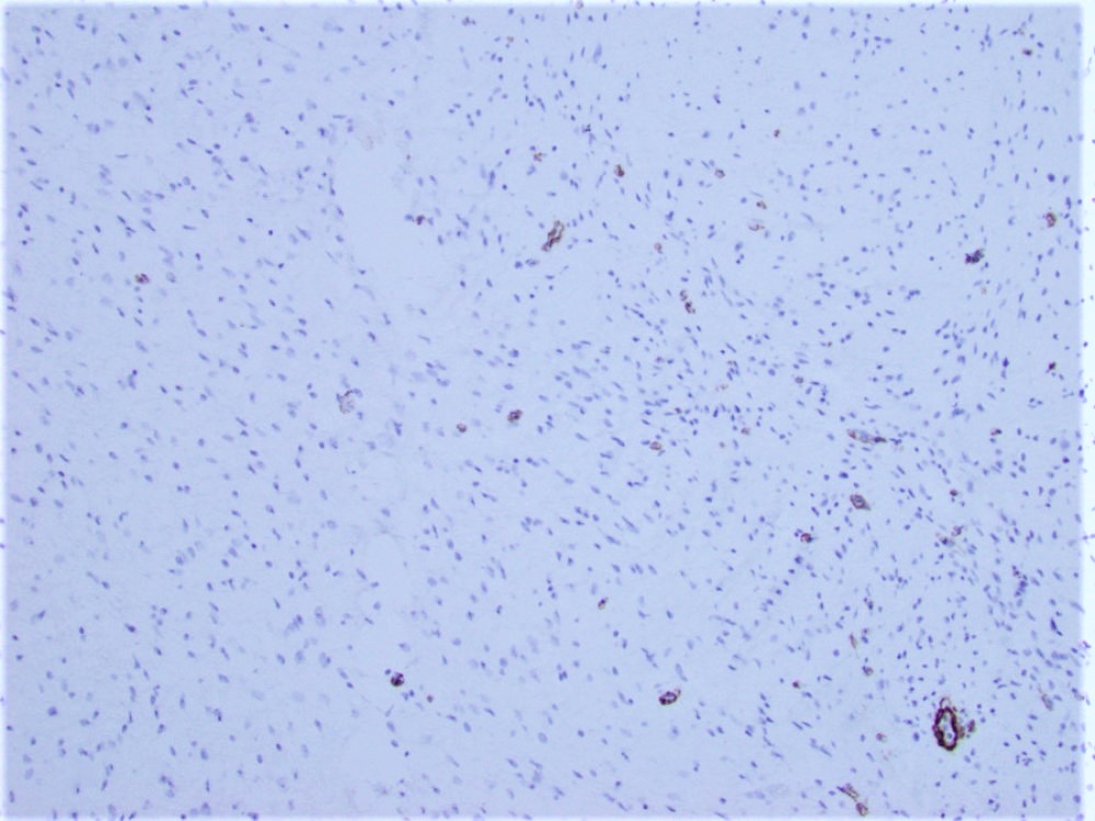
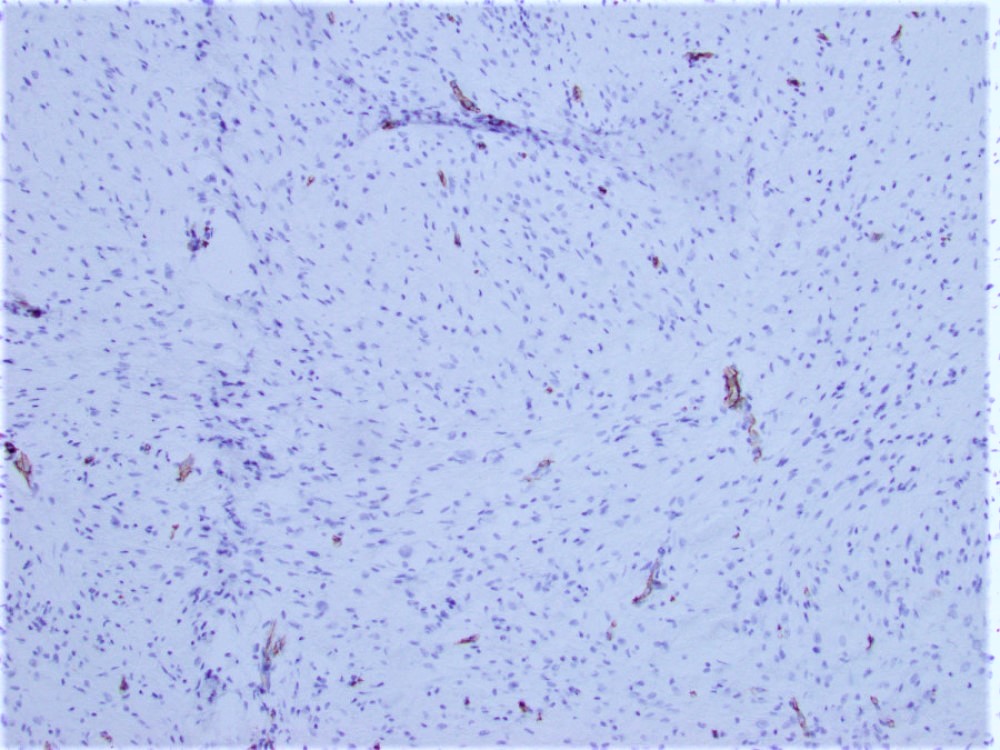
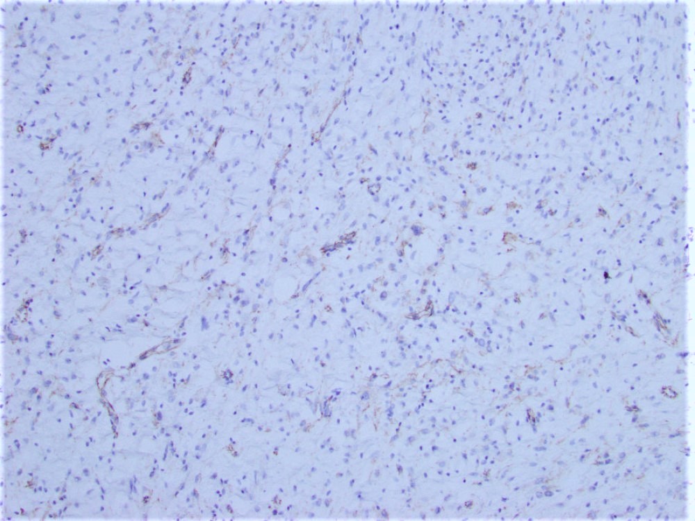
Sections of the biopsy revealed a proliferation of bland spindle cells arranged primarily in broad sweeping fascicles embedded in a dense collagenous stroma (Figure 2-3). There was no cytologic atypia and no mitoses were seen. Immunohistochemical staining demonstrated that the neoplastic cells were negative for SMA (Figure 4), desmin, CD34 (Figure 5), S-100 protein, and nuclear expression of β-catenin (Figure 6).