Detection of Antibody/Antigen Reactions by Immunoelectron Microscopy Using Protein A Gold
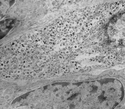
This technique allows the investigator to identify antibody/antigen complexes that localize to a particular subcellular organelle or compartment by using a preembedding streptavidin-biotin technique with diaminobenzidine (DAB) as a chromagen which is silver enhanced. The tissue is post-fixed in osmium tetroxide, dehydrated and embedded in EPON/Araldite resin.
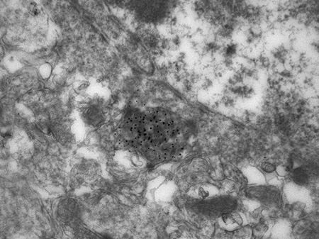
Protein A gold labeling of a prostatic endocrine-paracrine cell demonstrating localization of calcitonin (inset) to the neuroscretory granules.
Paraffin embedded tissue (same tissue) sections are cut and screened by light microscopy using a streptavidin-biotin technique to determine the appropriate antibody concentration to be used for the EM immunogold procedure. Ultrathin sections from the same block(s) are cut, incubated with primary antibody and then later incubated with Protein A gold particles (size range is 5 nm to 20 nm). The gold particles bind to the Fc portion of the antibody and are detected by EM. A variation of the large block "pop-off" technique for immunoelectron microscopy is also available.
Ref: de Mesy Jensen (Bentley), K.L. and di Sant'Agnese, P.A.: Large block embedding and "pop-off" technique for immunoelectron microscopy with applications to prostatic endocrine-paracrine cells. Ultrastruc. Pathol. 16:51, 1992.