

News
Muthukrishnan Earns $2.7M NIH Grant to Study Immune Dysfunction in Bone Infection
Tuesday, June 23, 2026
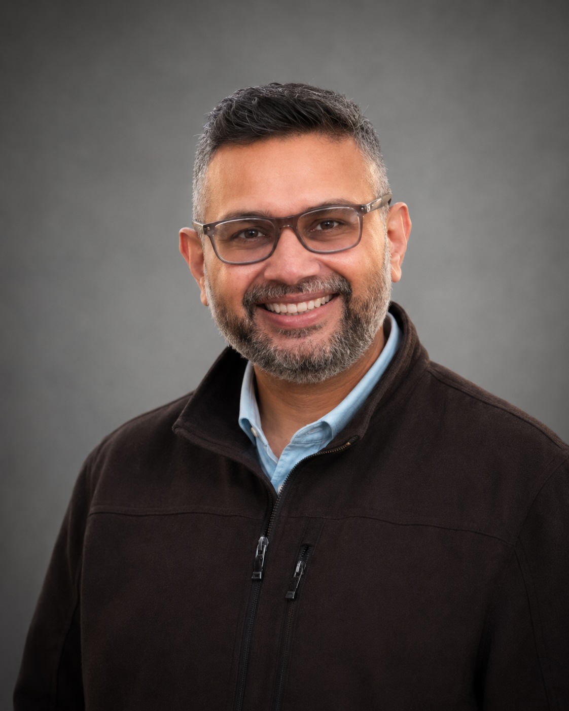
Gowri Muthukrishnan, PhD, assistant professor in the departments of Orthopaedics and Microbiology and Immunology, has earned a $2.7 million grant from NIH to study potential diagnostic and treatment options for implant-associated bone infection.
They are one of the most serious complications in orthopaedic surgery, with Staphylococcus aureus the leading cause of persistent cases. Despite advances in surgery and antibiotic therapy, recurrent and persistent S. aureus infections remain a major challenge in orthopaedic care, and reinfection rates following orthopaedic surgery have changed little over the past several decades. There are no evidence-based clinical assessment tools to evaluate severity of bone infection that could aid clinicians in patient care. At the same time, some patients recover from asymptomatic cases on their own; Muthukrishnan and other researchers are interested in how their immune systems effectively fight the infection, as it may lead to potential new prophylactic approaches or treatments.
The NIH grant will support Muthukrishnan’s research to better understand the mechanics of implant-associated bone infection; seek a host immune biomarker that could aid clinicians predict disease severity and treatment outcomes; and explore whether drugs now used in cancer treatment could help fight bacterial bone infection.
In prior research aimed at developing clinical biomarkers, Muthukrishnan’s lab performed host studies in a humanized mouse model and found that implant-associated osteomyelitis is characterized by increases in bacterial load and bone osteolysis, along with large numbers of proliferating human T cells adjacent to purulent abscesses in the bone marrow. The high levels of immune checkpoint proteins in patients with prosthetic joint infections were particularly apparent in patients with poor outcomes, including amputation due to persistent infection and septic death.
The work confirmed that during bone infections, there is dysregulated immune function; T cells experience “exhaustion” due to constant antigenic stimulation from bacteria, which prevents them from performing their intended immune functions effectively. Muthukrishnan’s research showed that serum immune checkpoint protein levels could predict patient outcomes with 90 percent accuracy, which could help orthopedic surgeons determine whether patients should receive aggressive or conservative treatment for joint infections. A biomarker could signal the severity of infection and shed light on why some patients’ bodies fight the infection successfully while others suffer devastating complications, including arthrodesis, reinfection, amputation, and septic death.
As part of the project, Muthukrishnan will assess patients based on their T cell exhaustion profiles. The NIH award will support one of the first comprehensive investigations into T cell exhaustion as both a biomarker and therapeutic target in orthopaedic infection. Immune checkpoint pathways triggered in cancer are also activated in bone infections; the research will explore whether immune checkpoint blockade drugs approved for cancer treatment could mitigate the infection burden and reprogram exhausted T cells, restore antibacterial immunity, and improve infection control. There are more than 2600 ongoing clinical trials for the drugs, but very limited research to date on their use for bacterial infection.
As part of the project, Muthukrishnan will collaborate with orthopedic surgeons to collect bone marrow samples during revision surgeries to analyze T cell exhaustion phenotypes.
“We hypothesize that persistent S. aureus infections cause impairment of CD4+ Th1/Th17 cells and can be leveraged as a functional biomarker of S. aureus implant-associated osteomyelitis disease outcome,” Muthukrishnan said.
Ultimately, Muthukrishnan aims to develop a simple blood test to help determine whether patients should receive aggressive or conservative treatment, and whether the FDA-approved checkpoint blockade drugs could become a standard of care for bone infections.
NIH S10 Award Will Expand Advanced Imaging Capabilities Across URochester Medicine
Wednesday, June 17, 2026
Published in the Center for Advanced Research Techologies (CART) Newsletter
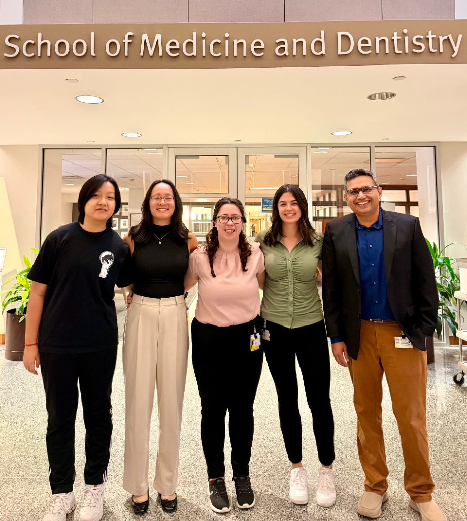
The University of Rochester has received an NIH Shared Instrumentation Grant (S10) to acquire a Bruker SkyScan 1276 CMOS Edition High-Resolution In Vivo MicroCT System, led by Hani Awad, PhD, Professor of Orthopaedics and member of the Center for Musculoskeletal Research. The approximately $466,000 award will support the purchase of a next-generation imaging platform that will be housed within the Biomechanics and Multimodal Tissue Imaging Core, replacing an aging scanner and significantly enhancing the institution’s capacity for high-resolution, longitudinal imaging studies. The system offers true spatial resolution down to 6 μm and advanced physiological monitoring capabilities, enabling researchers to visualize biological structures and disease processes with unprecedented detail.
The new microCT system will serve a broad community of NIH-funded investigators across University of Rochester Medicine, supporting research in musculoskeletal biology, cancer, cardiovascular disease, immunology, oral and craniofacial sciences, regenerative medicine, and other fields. Expected to support more than 1,600 hours of annual use, the instrument represents a significant investment in shared research infrastructure that will accelerate discovery, strengthen cross-disciplinary collaborations, and expand opportunities for translational research. This award reflects the collaborative spirit that drives scientific innovation at Rochester and highlights the impact of shared resources in advancing research across the institution.
TraCe-bMPS: Advancing Human Tissue-on-Chip Models for Discovery, Translation, and Drug Development
Wednesday, June 17, 2026
Published in the Center for Advanced Research Techologies (CART) Newsletter
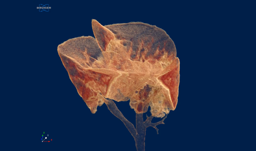
The Translational Center for Barrier Microphysiological Systems (TraCe-bMPS) is a consortium of industry and academia that is developing and applying chip-based models of human tissue for both discovery and regulatory drug development. Based at the University of Rochester, TraCe-bMPS grew out of collaborations between bioengineering labs with complementary strengths in materials, bio-sensing, and disease modeling. These teams created uniquely capable technologies for building and measuring compartmentalized models of human tissue. In TraCe-bMPS, the technology development teams are joined by regulatory and pharmaceutical scientists who bring quality systems, SOP development, and the reproducibility standards needed for the chips to support clinical drug development programs.
TraCe-bMPS teams are using the center’s tissue-on-chip platforms to model a range of healthy and diseased tissues in a number of contexts. Current efforts include models of neurovascular injury, inflammatory arthritis, connective tissue repair, retinal barrier function, and innate immunity. Some tissue chips are developed in collaboration with pharmaceutical and academic partners with specific clinical and preclinical endpoints in mind. Others are used as platforms for discovery and hypothesis testing about the nature of multicellular interactions in disease origins and progression.
The adoption of human microphysiological systems and other non-animal methodologies (NAMS) is being driven by federal mandates and an ever-growing interest in the reduction of animal use in biomedical research and drug development. TraCe-bMPS is a new type of core facility that is appearing at universities to meet the need for rapid adoption of NAMS by biomedical research programs. TraCe-bMPS has packaged its core capabilities as services to help teams internal and external to Rochester adopt NAMS. The center operates a service core providing quality-controlled chip components, validated protocols, and technical support to investigators across universities, medical centers, and industry labs. To date, more than 20,000 devices have been built and used in research laboratories around the world. TraCe-bMPS also supports adoption through a training program at Rochester, and external workshops.
If interested in learning more, contact Jim McGrath or visit the website →
Calvin Cole, Zach Sechrist: Mentorship That Shapes the Whole Scientist
Tuesday, April 28, 2026
From the Faculty Focus Newsletter, Education Update
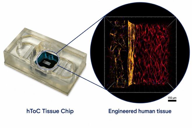
In this episode of Learners on Location, the assistant professor of Surgical Oncology and the fifth-year Pathology PhD candidate demonstrate how mentorship goes far beyond the lab. Their approach, grounded in wellness, growth, and purpose, demonstrate how personal wellbeing can lead to meaningful scientific impact.
Where's Wilmot's Director: Exploring the Cancer Microenvironment with Dr. Yeh's Lab
Friday, April 10, 2026
In April's installment of "Where's Wilmot's Director?", Dr. Jonathan Friedberg visted Shu-Chi (Allison) Yeh's lab in the CMSR. Dr. Friedberg met with Dr. Yeh and her multidisciplinary team to learn more about their insights and use of innovative technology in investigating the cancer microenvironment, a reasearch area of strength at Wilmot with new translational opportunities.
Click here to read more about Dr. Friedberg's visit to the Yeh Lab.
Gregg Nicandri Tapped to Lead New Digital Strategy & Innovation Office
Friday, March 20, 2026
URMC CEO David Linehan has named UR Medicine’s Chief Medical Information Officer Gregg Nicandri, MD, to lead the office as its inaugural Chief Digital & Innovation Officer (CDIO), charged with advancing an enterprise digital strategy to ensure innovation and execution are tightly aligned and positioned to deliver meaningful impact.
“We’re not running to catch up but leading the future,” said the new Chief Digital and Innovation Officer, who will work to advance an enterprise digital strategy, moving quickly and with intention to innovate and execute for meaningful impact. Linehan noted Nicandri’s experience has prepared him to forge our digital future. “He has worked tirelessly to develop innovative provider enablement tools to improve wellbeing by freeing up the time spent on administrative tasks to allow more quality face-to-face interactions with patients,” Linehan said.
Read about Dr. Nicandri's plans to propel the digital future of URMC.
Collaborative Effort Expands Upon Study Linking Taurine to Cancer
Friday, January 23, 2026
In a groundbreaking basic science study last year, researchers discovered that taurine, available in energy drinks and as a supplement, feeds the growth of leukemia stem cells. A new study recently published by the same Wilmot Cancer Institute team expands that work and suggests that as taurine fuels leukemia, it becomes less available for other normal cells and may result in weaker bones in mice.
The bone marrow microenvironment is key to the latest research from the Bajaj lab, and has been a longtime focus of scientists who are members of Wilmot’s Cancer Microenvironment (CM) research program. Not only do many types of cancer, including leukemia, develop in the bone marrow, but the interactions and signaling between all cells and tissues in the bone marrow environment are critical for normal bone and blood development, while also supporting cancer.
PhD candidate Christina M. Kaszuba, a student in the University of Rochester Biomedical Engineering graduate program, led this project. She works exclusively in the Bajaj lab studying cells and components of the bone that contribute to cancer development, with the goal of finding new cancer drugs.
Bajaj is an assistant professor of Biomedical Genetics and a recipient of an American Society of Hematology Scholar Award and Leukemia Research Foundation New Investigator Award.
The research, funded by the National Institutes of Health (NH) is the result of a broad collaboration that also includes Laura Calvi, MD, professor of Medicine and co-director of the CM research program; Jane Liesveld, MD, professor of Medicine; Hani Awad, PhD, professor of Orthopedics; Roman Eliseev, MD, PhD, associate professor of Orthopedics; and teams at the University of Rochester Center for Musculoskeletal Research and Wilmot Shared Resources.