Case of the Month: 78-Year-Old Male with Left Renal Mass
By Sohaib Abu-Farsakh, MD (PGY-4)
Clinical history:
A 78-year-old male presented with gross hematuria. Past medical history includes prostate cancer and mitral and aortic valve replacement.
Recent history:
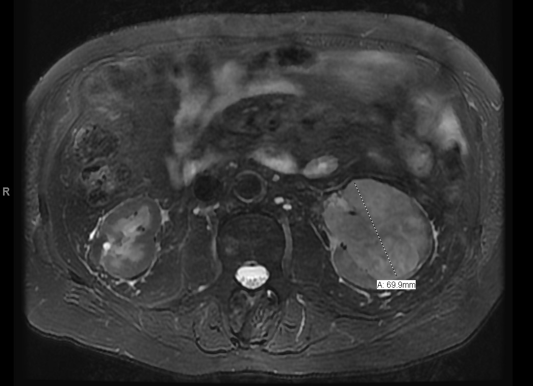
CT scan revealed a left renal mass. MR imaging of the abdomen revealed a 7 cm infiltrating left-sided renal mass which extended into the collecting system and proximal ureter (Fig. 1). Also seen was a 3.7 cm. enhancing, heterogeneous soft tissue mass involving the renal pelvis and proximal ureter.
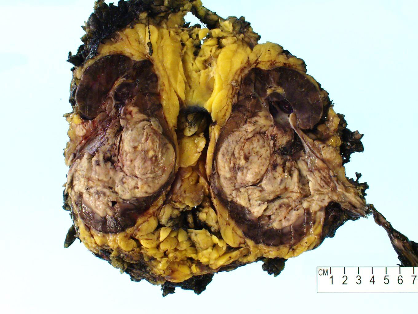
Left renal artery embolization was done to control the hematuria and the patient underwent left radical nephrectomy. Sectioning of the nephrectomy specimen revealed an 8.5 x 6.5 cm. tan-white heterogenous necrotic mass that replaced approximately 80% of the kidney and extends to the hilar fat (Fig. 2). Also seen was a 4 cm firm mass with tan-white cut surfaces present in the proximal ureter.
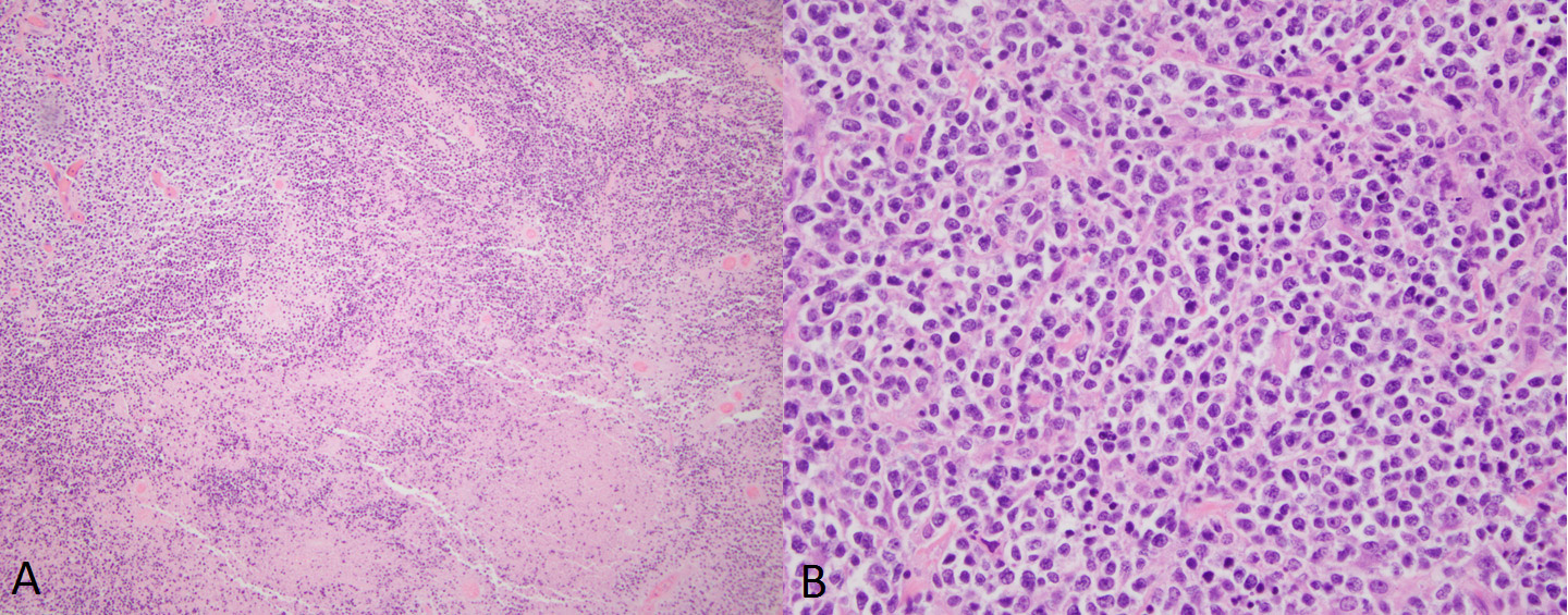
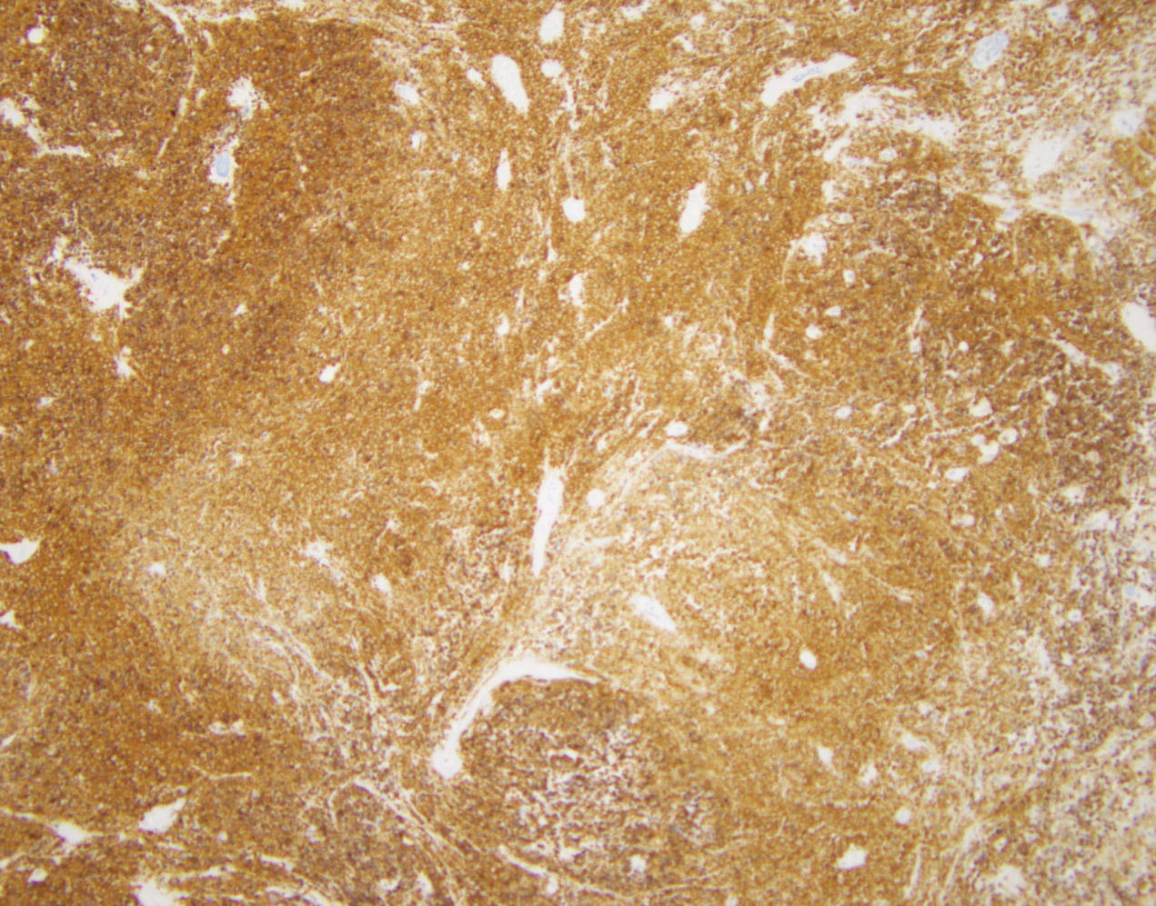
Histologic sections revealed replacement of the renal parenchyma by an infiltrate of small round blue cells with wide areas of necrosis (Fig. 3A). The infiltrating cells are pleomorphic with high nuclear/cytoplasmic ratio (Fig. 3B). Frequent mitotic figures and apoptotic bodies were identified. Immunostaining for CD20 showed diffuse wall-to-wall positivity (Fig. 4).
Additional immunostaining was performed and is shown in the table below:
| Positive Stains | Negative Stains |
| CD45 (leukocyte common antigen) | CD3 |
| CD20 | CD5 |
| BCL-2 | PAX-8 |
| BCL-6 | GATA-3 |
| CD10 | MUM1 |
| Ki67-up to 90% | C-MYC |
| TdT | |
| Cyclin-D1 |