Case 76: Lytic Bone Lesions
Roula Katerji, MD and Aaron R. Huber, DO
Past Medical History
A 70-year-old patient with a history of lung adenocarcinoma
Recent History
The patient presented with right lower leg pain. Computed tomography scan of the femur showed lytic lesions with soft tissue extension.
Pathology
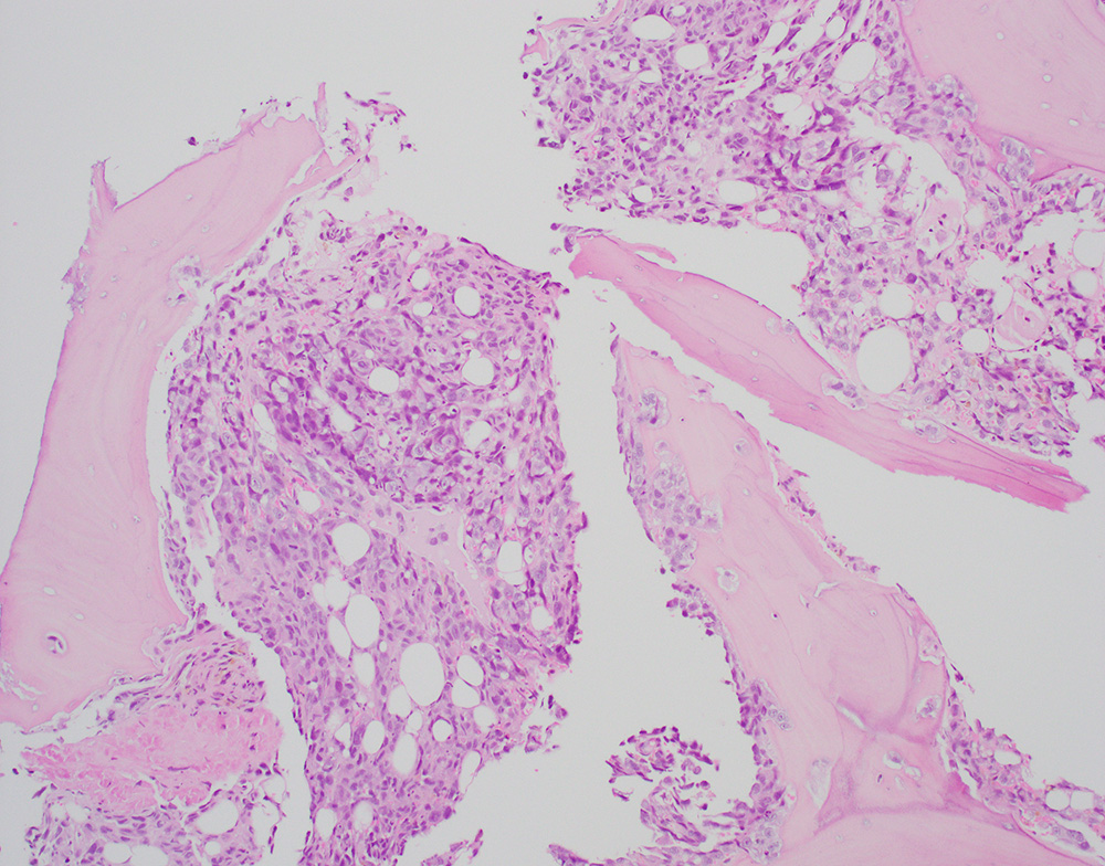
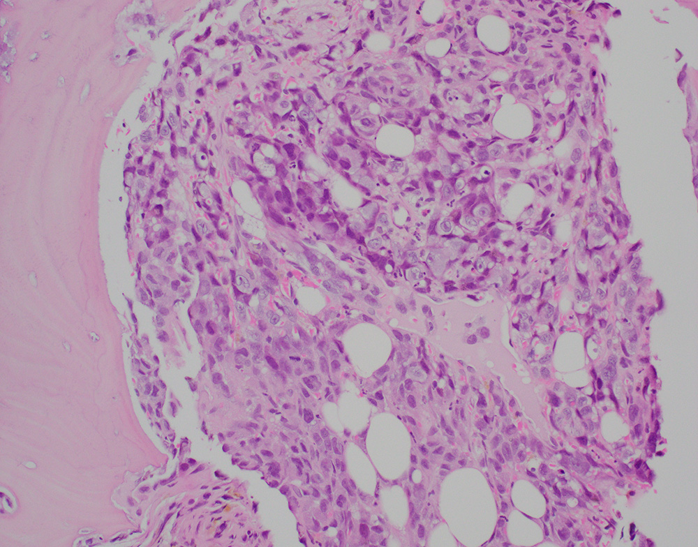
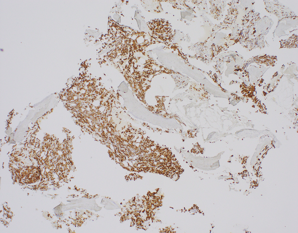
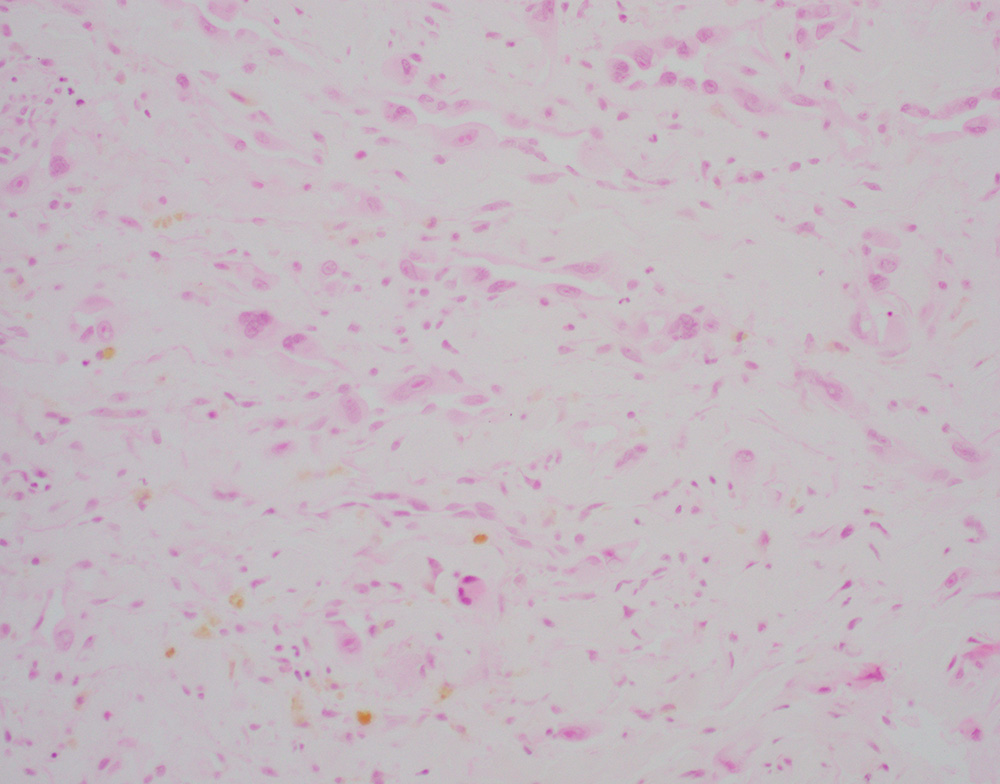
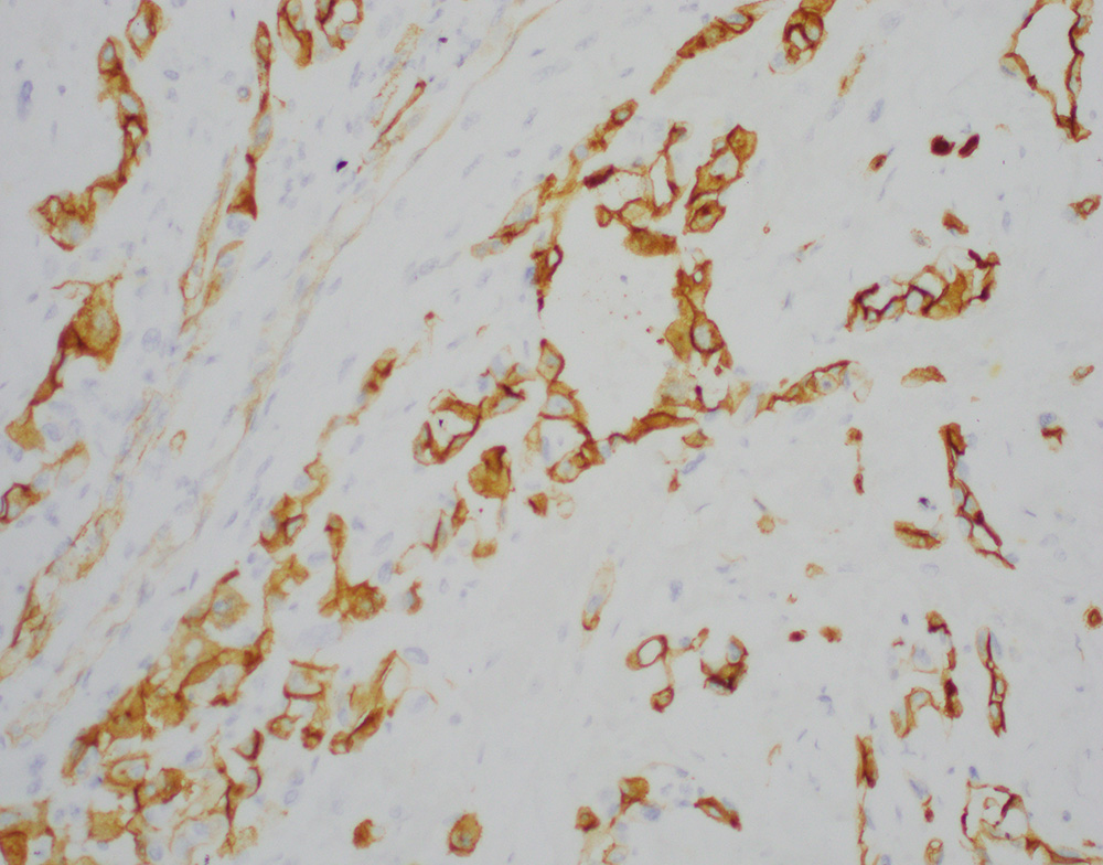
A bone biopsy showed a poorly differentiated epithelioid neoplasm with pleomorphic nuclei, prominent nucleoli (Figures-1, 2) with foci of necrosis. The tumor cells were positive for keratin 7 (Figure-3) with patchy positivity for pan-keratin, while they were negative for PAX8, SOX10, keratin 20, TTF-1, p40, NKX3.1, CDX2, and GATA3. Given the keratin positivity, the tumor was felt to represent metastatic carcinoma. The patient also had several skin nodules involving the posterior leg. A skin biopsy demonstrated numerous irregularly shaped anastomosing and infiltrative vascular channels lined by atypical endothelial cells (Figure-4). The neoplastic cells were positive for ERG, CD31 and CD34 (Figure 5). Re-evaluating and staining the bone biopsy with vascular markers showed positive ERG staining in the neoplastic cells.