Case of the Month: Abdominal Mass
By Kingsley Nwokelo, MS3
Clinical history
A 58-year-old woman with a one week history of constant mid to right lower quadrant abdominal pain. Associated with early satiety, loss of appetite, and increased urinary frequency.
Past medical history
Past medical history is significant for hypertension, GERD, sarcoidosis, and prior hysterectomy.
Recent history
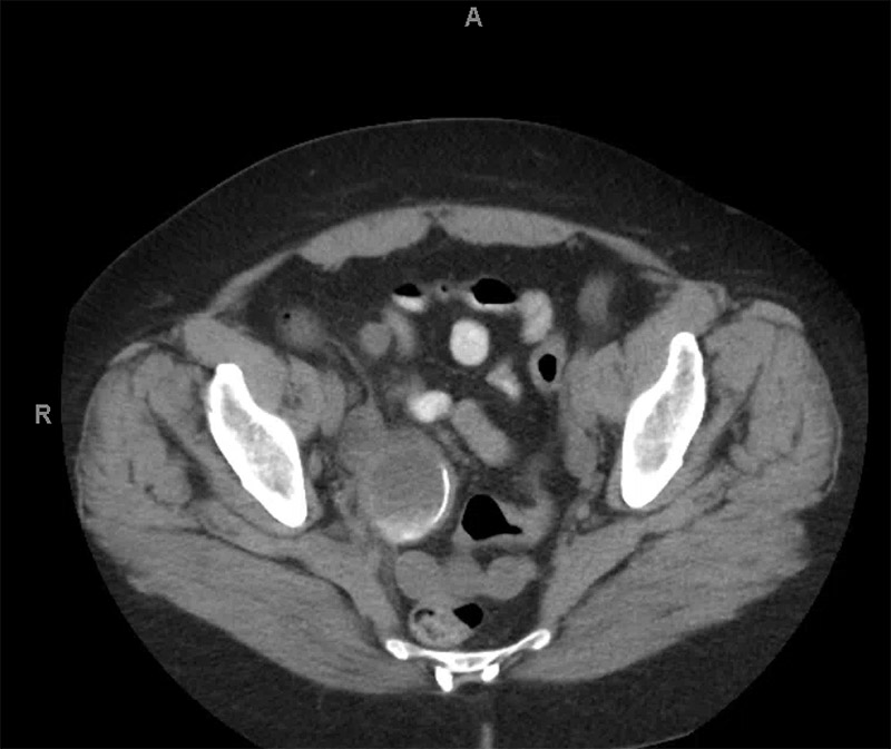
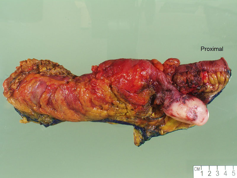
Abdominal CT scan demonstrated a distended appendix with peripheral and dependent calcifications (Figure 1). Right hemicolectomy with ileocolonic anastomosis was performed. Gross examination of the right hemicolectomy specimen included a 6 cm tan-white and firm mass involving the distal appendix (Figure 2). The lumen of the appendix communicated with the central portion of the tumor, both of which contained mucinous material.
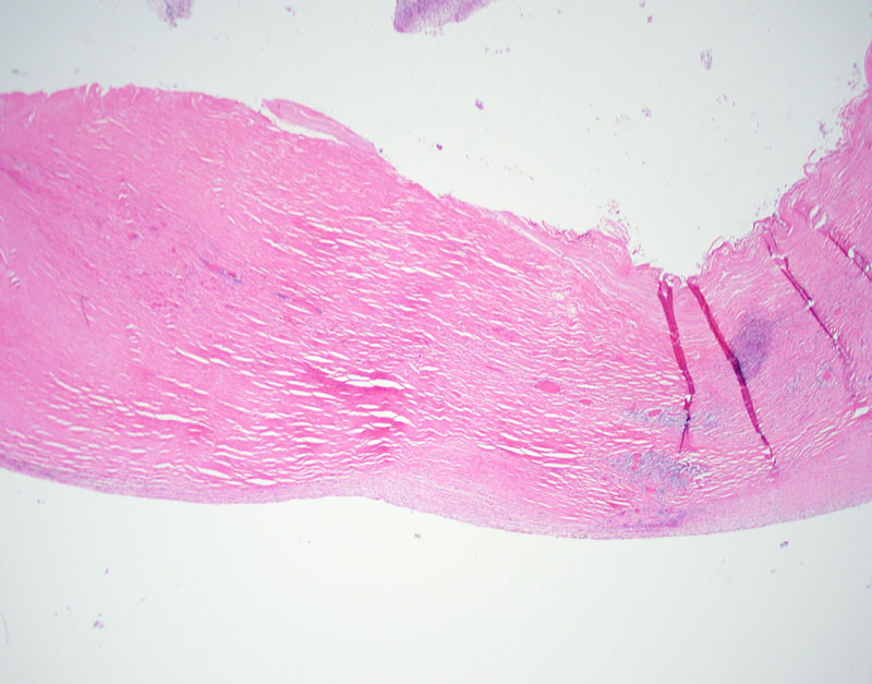
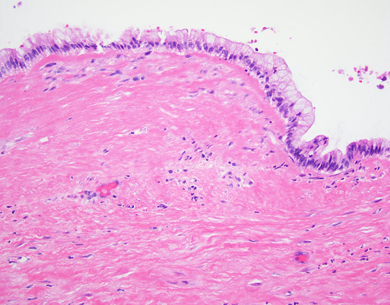
Microscopic examination of the mass demonstrated a thickened and fibrotic wall with focal acellular mural mucin and calcifications (Figure 3). The majority of the luminal surface was denuded, however focal areas demonstrated mucinous epithelium (Figure 4).