Case of the Month: Migrating Abdominal Pain
By Andrew Soroka, MD
Clinical History
A previously healthy 69-year-old woman presented with a 4 month history of migrating abdominal pain. The pain started in her right shoulder, then migrated to her left flank, and then to the right upper quadrant and the right axilla.
Past Medical History
No significant past medical history was noted.
Recent History
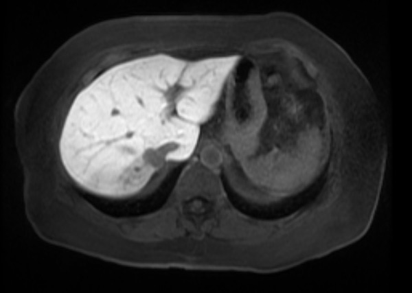
MRI with and without contrast showed a 2.0 x 1.6 cm mass in the posterior right hepatic lobe, immediately posterior to and contacting the IVC (Figure 1). The patient subsequently underwent a right hepatectomy, including a small portion of the IVC.
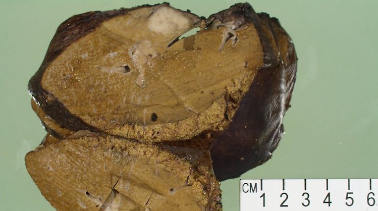
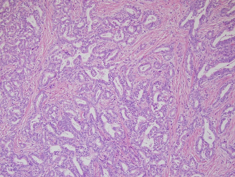
Grossly, the right hepatectomy specimen weighed 698 grams and cut surfaces demonstrated a 2.9 x 2.1 x 1.2 cm firm, pale, white mass with an apparent tumor thrombus (Figure 2). Histologically, the tumor was composed of tubules within a densely sclerotic and desmoplastic stroma (Figure 3). An elastin stain was performed which highlighted venous invasion. An immunohistochemical stain for HepPar-1 and a Kreyberg mucin stain were negative.