Case of the Month: Painless Nodule of the Breast
By Cynthia Reyes-Barron, MD
Clinical History
An elderly female presented with a persistent 0.5 cm, slightly firm, red, painless nodule involving the skin of the left breast.
Past Medical History
Eight years prior to the development of the skin nodule , the patient was diagnosed with 1.1 cm infiltrating ductal carcinoma (IDC) of the left breast (nuclear grade 2) that was estrogen receptor (ER) positive, progesterone receptor (PR) negative, and human epidermal growth factor receptor 2 (HER2) negative. She was treated with a partial mastectomy, whole breast radiation therapy, and an aromatase inhibitor.
Five years after the initial breast carcinoma, she underwent a second partial mastectomy of the left breast for recurrent IDC (nuclear grade 2), again ER positive, PR negative and HER2 negative. She received combination chemotherapy with doxorubicin, cyclophosphamide, and paclitaxel followed by focused partial breast radiation therapy and treatment with an aromatase inhibitor.
Six months prior to the current biopsy specimen she presented with a 2 cm red nodule at the same site as the current lesion. A skin punch biopsy showed dermal fibrosis and scattered perivascular chronic inflammation.
Recent History
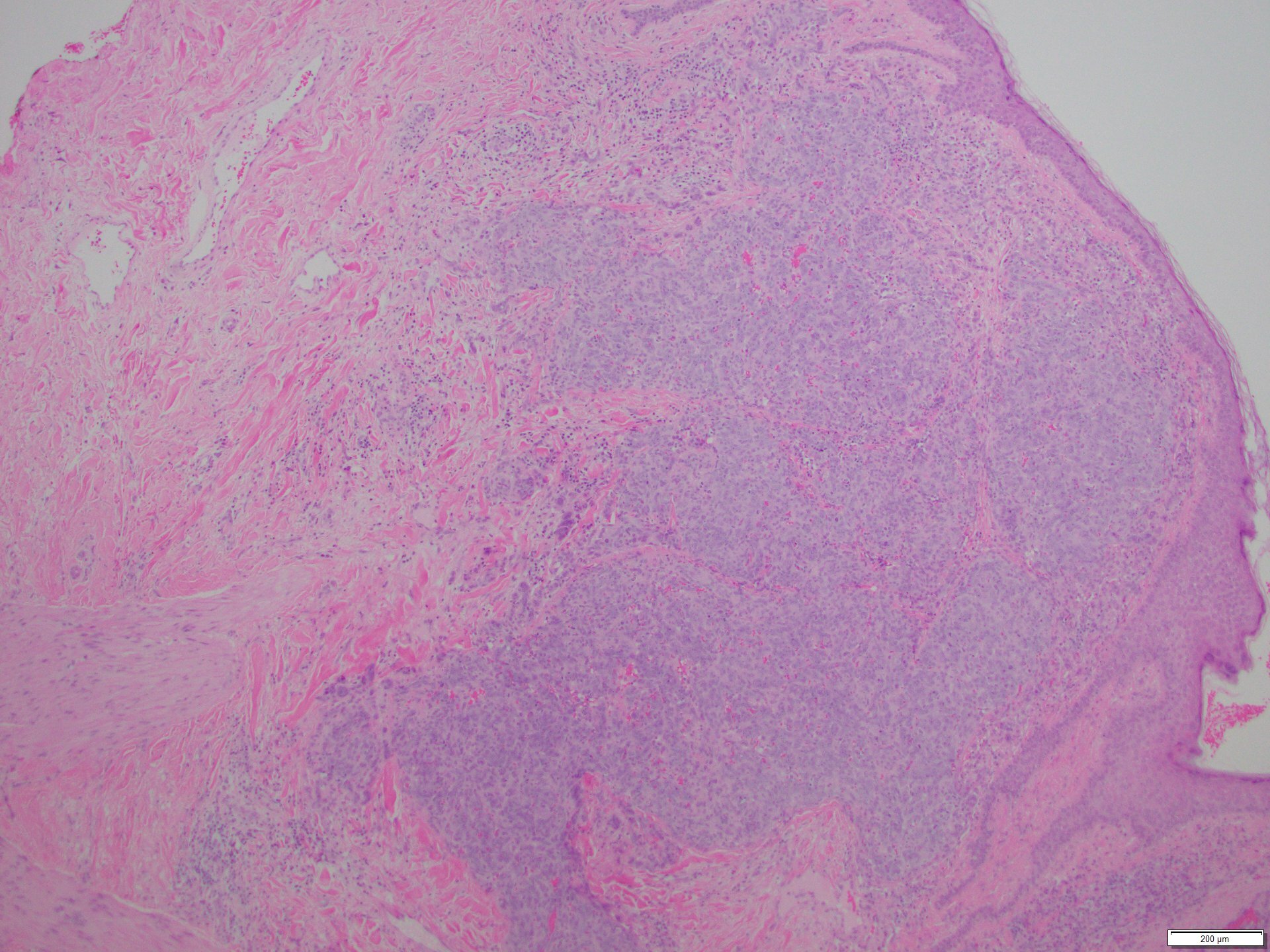
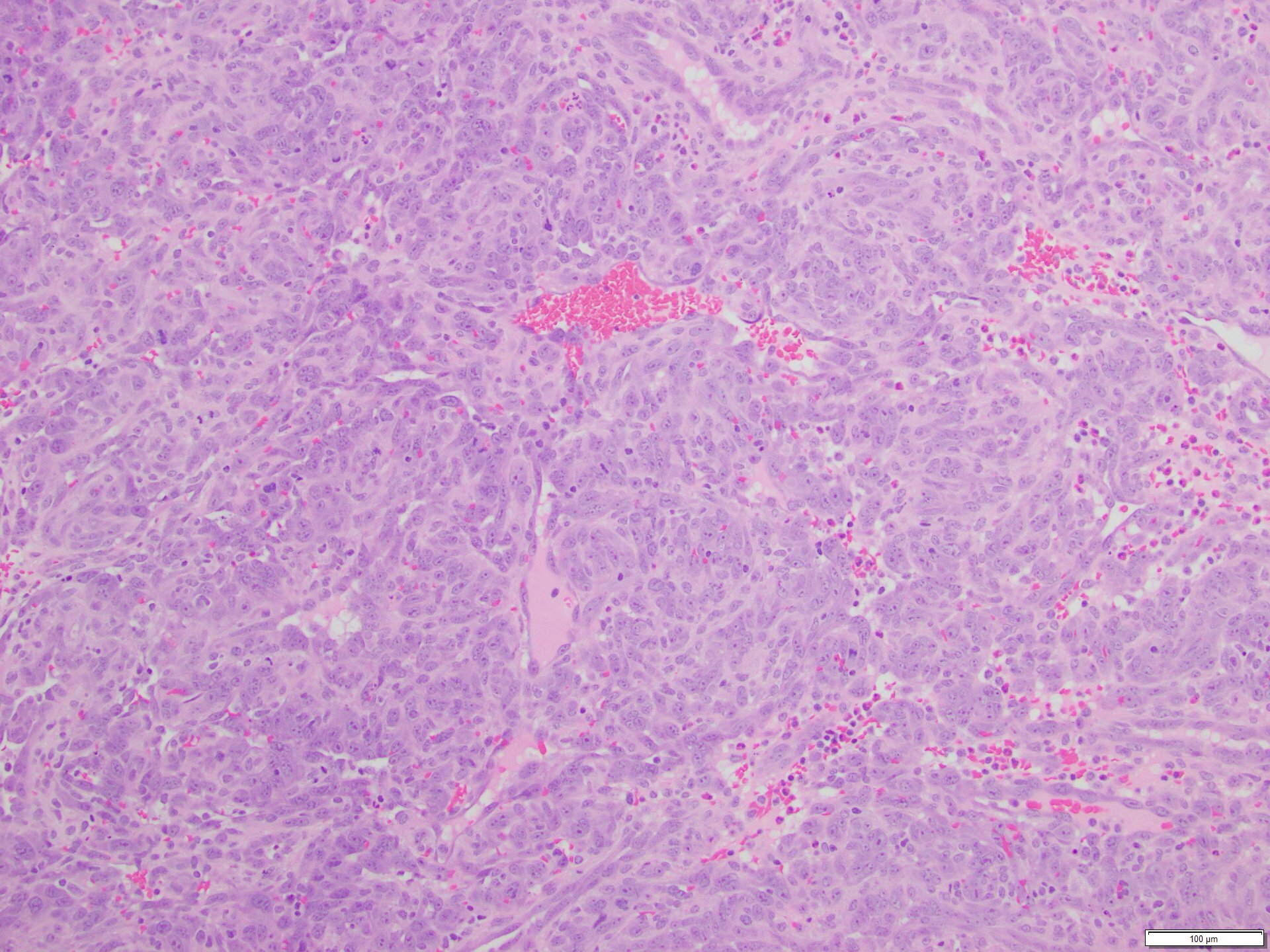
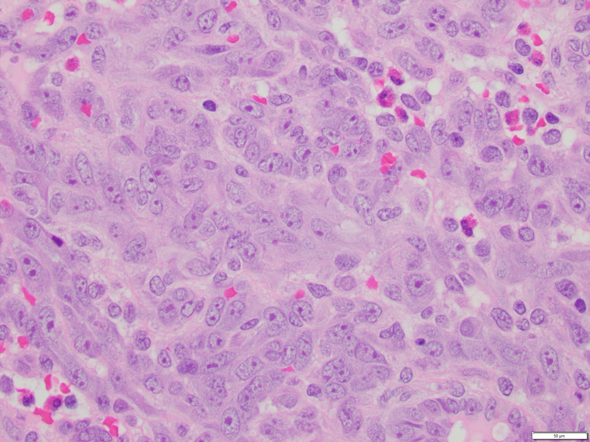
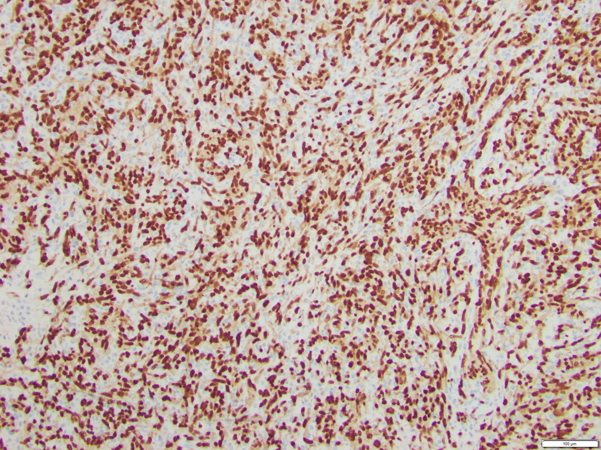
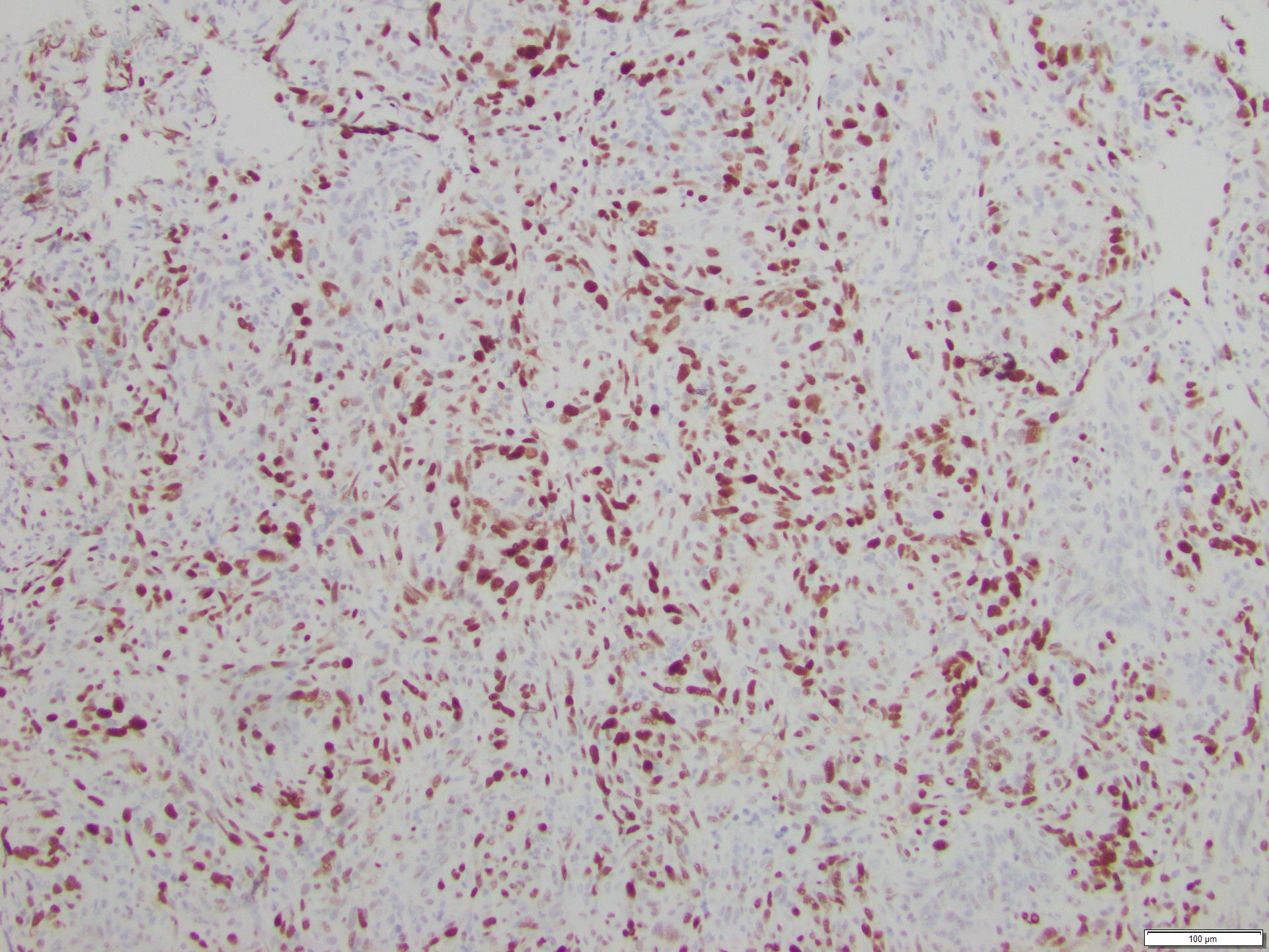
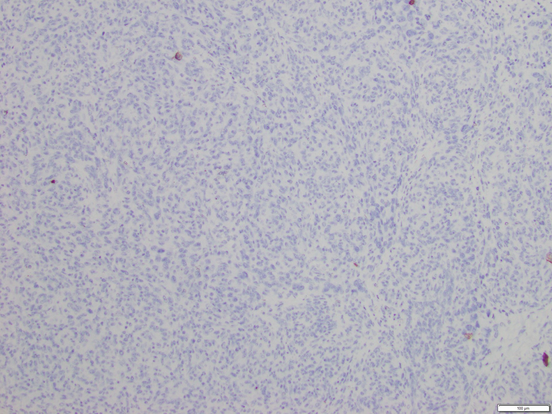
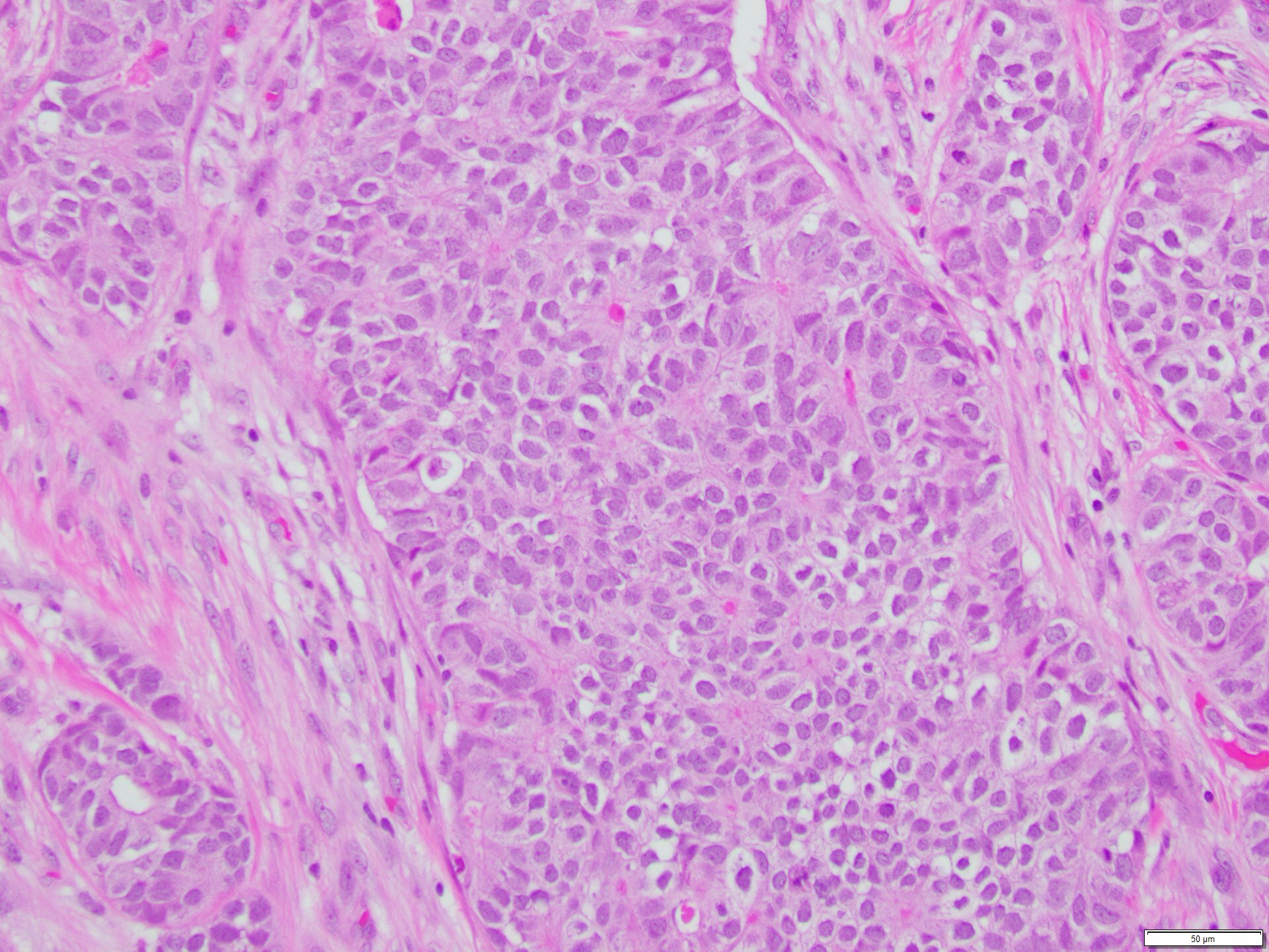
The skin lesion was completely excised and sent to pathology for diagnosis (Figures 1-3). The lesion is hypercellular with an infiltrative, predominantly solid, growth pattern in the dermis and subcutaneous tissue. It is highly vascular and has complex anastomosing and poorly formed vascular channels. The tumor cells are large, pleomorphic, have vesicular chromatin, and prominent nucleoli. There are frequent mitoses and extravasated red blood cells throughout. Immunohistochemistry shows the tumor cells are diffusely positive for ERG (Figure 4) and c-MYC (Figure 5) while negative for pan-cytokeratin (Figure 6) and GATA3. Subsequently, the patient underwent a left total mastectomy.