Case of the Month: Rapidly Enlarging Lung Mass
By Phoenix Bell, MD (PGY-3) and Moises Velez, MD
Clinical History
A 46-year-old female with a past medical history significant for invasive ductal carcinoma, status post lumpectomy and radiation, presents with an enlarging right upper lung lobe lesion.
Recent History
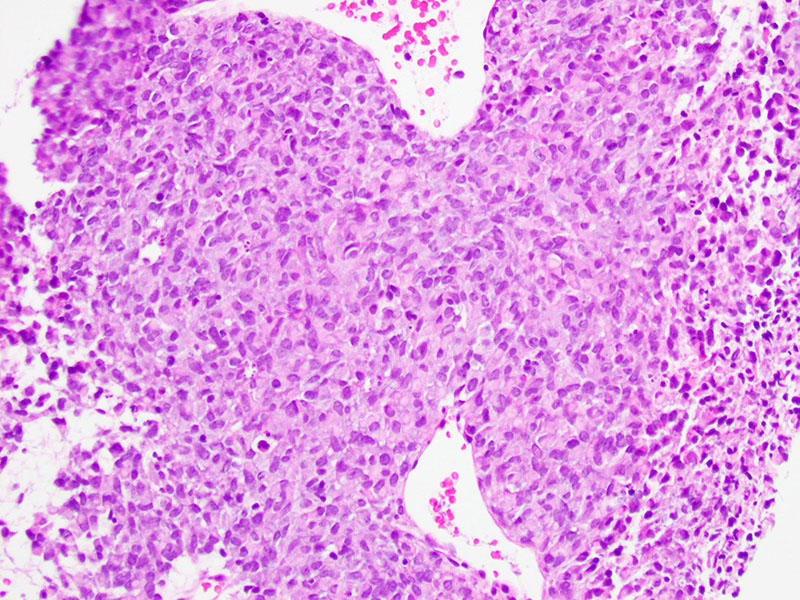
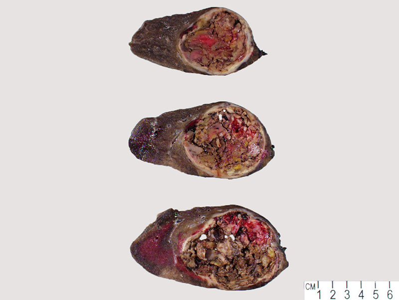
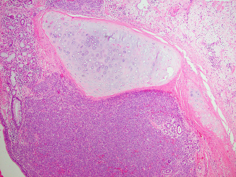
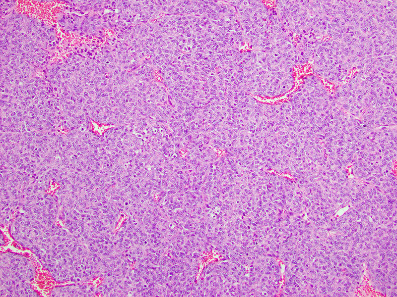
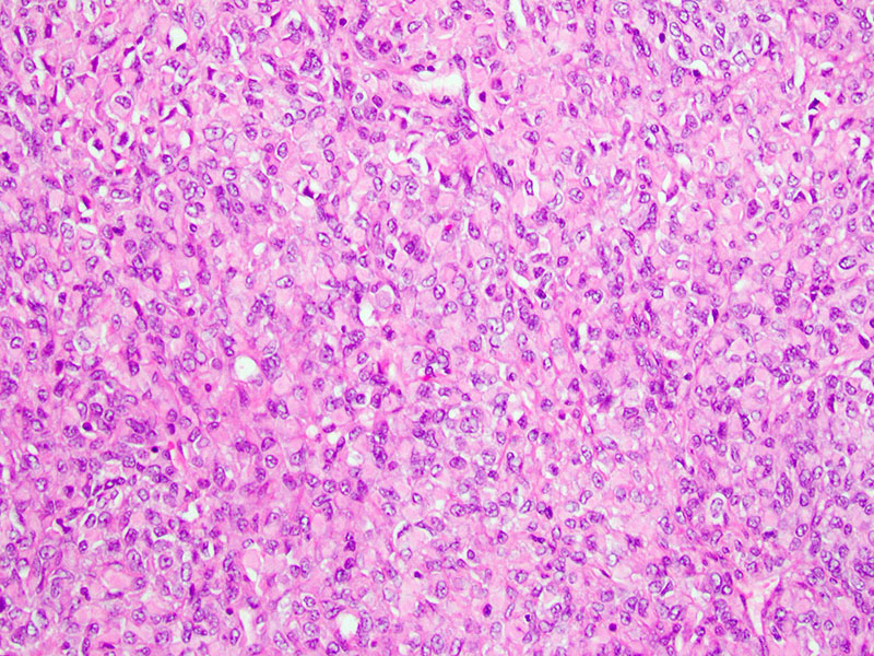
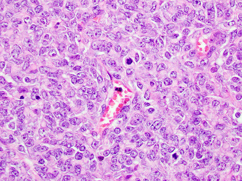
One year earlier, during an evaluation for her newly diagnosed breast cancer, a CT scan was ordered, which revealed a 2 cm right upper lung lobe lesion. The radiologic impression was that of a granuloma. One month ago, a follow-up PET scan showed FDG positivity in her bilateral ovaries and the right upper lobe lesion, which had grown to 10 cm. CT-guided biopsy of the mass revealed a monotonous population of malignant cells with eosinophilic cytoplasm and round to ovoid eccentric nuclei (Figure 1). Subsequently, the patient underwent a lobectomy. Gross examination of the right upper lobe demonstrated an 11.1 x 8.1 x 6.5 cm mass with a tan-brown to yellow, extensively friable, and hemorrhagic cut surface (Figure 2). Histologic examination showed sheets of epithelioid cells (Figure 3) surrounding scattered blood vessels (Figure 4). In some areas, the cells showed prominent cytoplasm with eccentric nuclei (Figure 5), as seen on the prior biopsy. Mitotic figures were readily identified (Figure 6). The tumor cells were positive for ER and vimentin, while negative for GATA3, CD68, Napsin A, TTF-1, MART1, PR, CD45, P63, CK5/6, and STAT6. INI-1 was retained and FISH was positive for the SYT gene rearrangement.