Case of the Month: Liver Masses
By Caroline Miller, M.D. (PGY-2) and Xiaoyan Liao, M.D., Ph.D.
Clinical history
A 55-year-old female has abnormal imaging results of the liver during routine surveillance.
Past medical history?
Past medical history was significant for choroidal melanoma of the right eye diagnosed several years ago. She received treatment with laser photocoagulation and intravitreal bevacizumab, underwent a barrier laser procedure and is receiving ongoing local radiation therapy via radioactive plaque implant. She was surveilled annually for metastasis with chest computed tomography scan, liver function tests and hepatic ultrasound.
Recent history
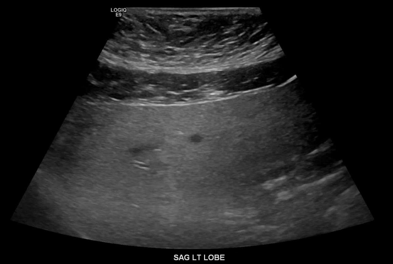
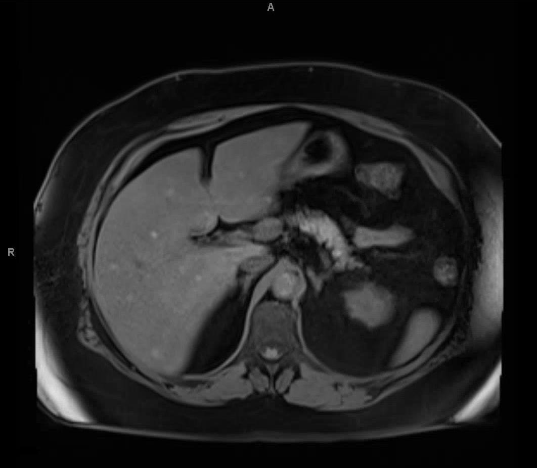
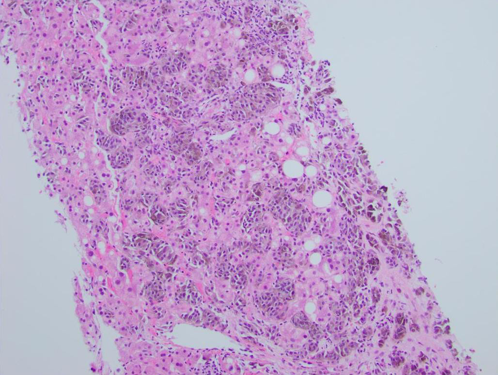
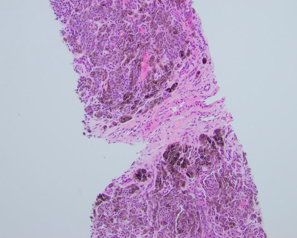
The patient underwent an abdominal ultrasound which revealed multiple hypoechoic lesions less than 1 cm dispersed throughout the liver (Figure 1). Magnetic resonance imaging was also performed and demonstrated multiple lesions dispersed throughout the liver (Figure 2). In addition, both imaging modalities showed a background of hepatic steatosis. Biopsy of a representative liver lesion demonstrated a malignant neoplasm infiltrating the hepatic parenchyma arranged primarily in a nested pattern (Figure 3). The neoplasm was composed of spindled and epithelioid cells with deep brown pigmentation within the cytoplasm (Figures 4 and 5). Given the morphologic findings, no immunohistochemical studies were performed.