Case of the Month: Lower Leg Skin Lesion
By Claire Porterfield, BS (Year Out Student Fellow), Xi Wang, MD and Glynis A. Scott, MD
Clinical History
An 88-year-old “Mediterranean” man presented with an exophytic, vascular-appearing nodule in the setting of purple plaques and patches on his lower legs. A biopsy of the lesion was performed, given the clinical concern for Kaposi sarcoma (KS).
Past Medical History
Stasis dermatitis, hypertension and Parkinson’s disease.
Histology
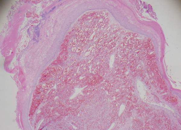
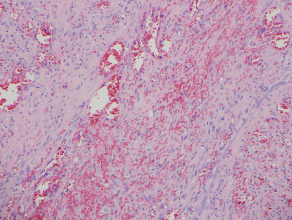
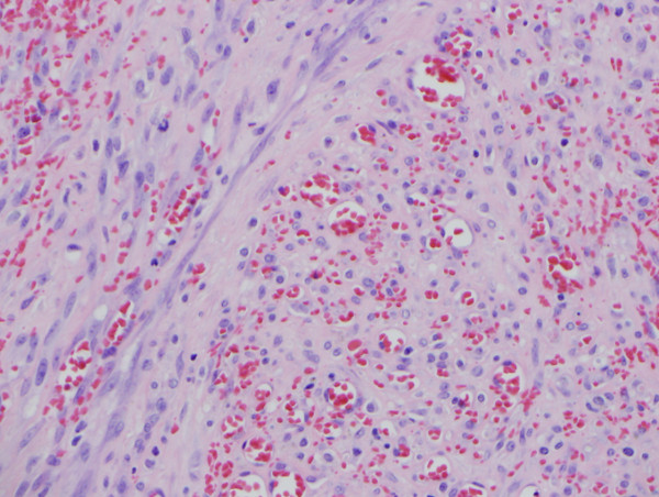
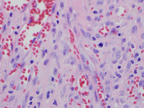
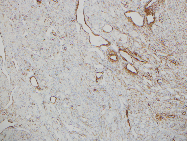
Histologic sections of the biopsy demonstrated a circumscribed tumor with areas of ulceration, consisting of a network of blood-filled spaces associated with a cellular spindle cell component (Figures 1 and 2). Cytologically, the cells lacked significant pleomorphism, but occasional mitotic figures were identified (Figure 3 and 4). Some cells showed a more epithelioid morphology. Human Herpes Virus 8 (HHV8) immunohistochemical stain was performed and was negative. A smooth muscle actin (SMA) immunohistochemical stain was positive (Figure 5).