Case of the Month: Bilateral Ovarian Masses
Authors: Harsimran Kaur, MBBS, MD and Sharlin Varghese, MBBS
Clinical History
A woman in her thirties presented with 2 years of progressively increasing size of abdomen and urinary frequency.
Past Medical History
The patient has been oligomenorrheic (1-2 menses/year) for the past 2 years and was on monthly oral contraceptives. Prior to that she had regular menses.
Recent History
The patient was noted to have a pelvic mass at annual well-woman exam. Computed tomography scan of the abdomen and pelvis with intravenous contrast demonstrated a large (16 x 12 x 11 cm) predominately solid and partly cystic mass originating from the left ovary. Differential diagnostic considerations, from a radiographic standpoint, included both benign and malignant neoplasms. The right ovary was noted to have a separate mature cystic teratoma (6 x 4.5 cm).
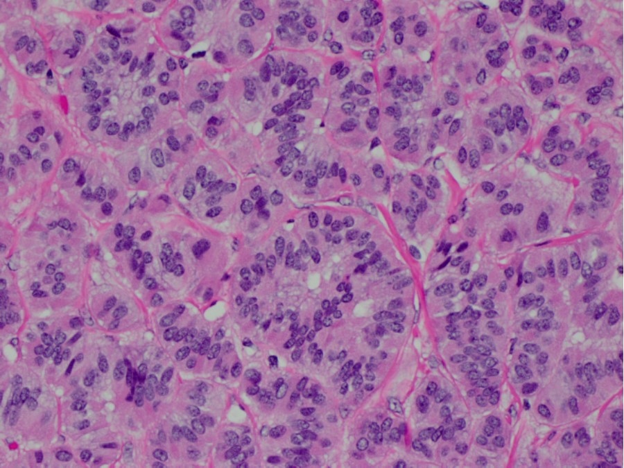
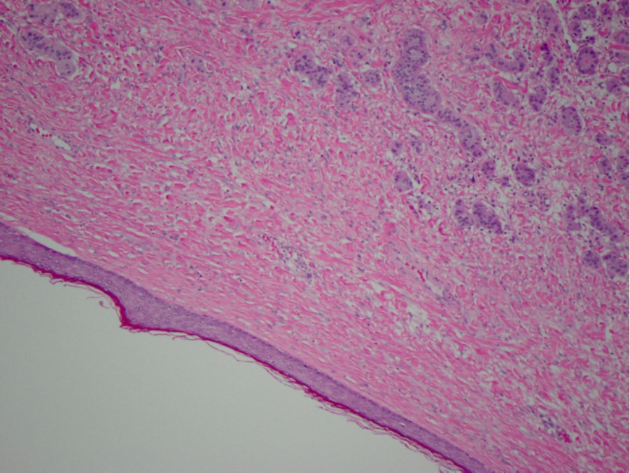
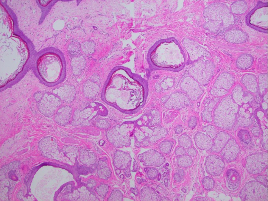
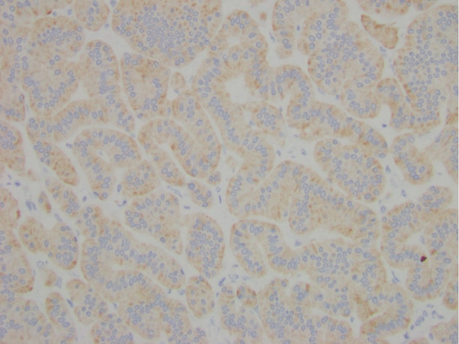
Histologic sections of the left ovary demonstrated parallel cords and ribbons of cells with bland, round nuclei and ample cytoplasm separated by fibrous stroma. (Fig. 1) associated with a mature teratomatous component (Fig 2). Histologic sections of the right ovary demonstrated a mature cytstic teratoma composed predominately of ectodermal components (Fig. 3). Immunohistochemical stains were performed and the cells of interest were positive for synaptophysin (Fig. 4), chromogranin and CD56 (Fig. 5). A proliferative index of less than 1% was seen with Ki-67 in the neoplastic cells.