Case of the Month: Hip Mass
Case Authors: Caroline Miller, MD and Aaron R. Huber, DO
Clinical History
A middle-aged woman presented with a hip mass identified on a work-up for leg pain.
Past Medical History
Significant for asthma, fibromyalgia, and gastroesophageal reflux disease.
Recent History
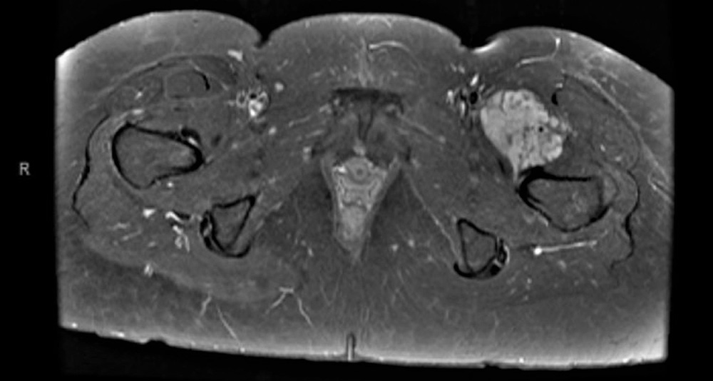
The patient had a one-year history of leg pain that had become more intense during the prior 2-3 months. Magnetic resonance imaging demonstrated a 7.4 cm in greatest dimension moderately hyperintense mass within the muscular compartment of the anterior left hip on T2-weighted images (Figure 1). The mass contained elements isointense with skeletal muscle and fat on T1-weighted images. The mass also contained blood vessels demonstrating flow-voids and significant enhancement on post-contrast images. The patient underwent an ultrasound-guided core needle biopsy of the mass.
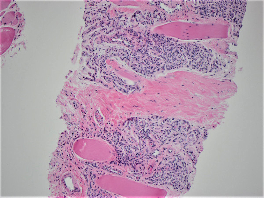
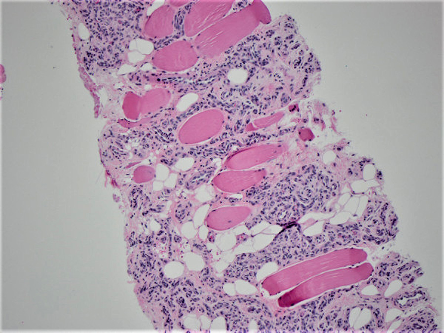
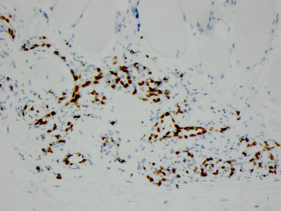
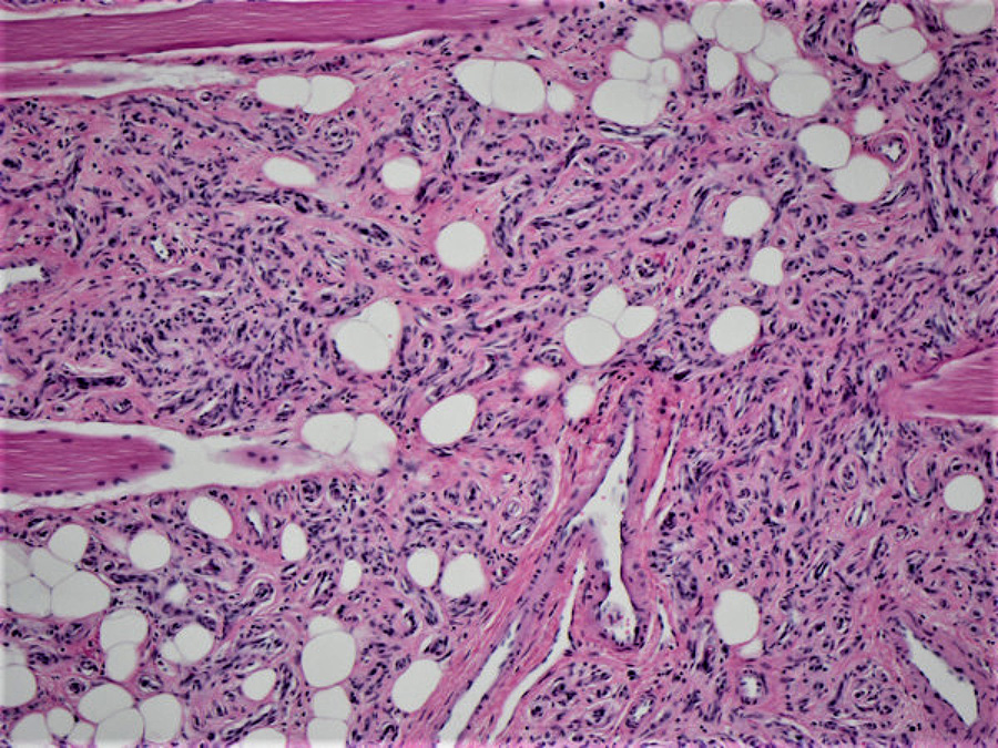
Histologically, the biopsy demonstrated numerous benign, variably-sized vascular spaces infiltrating the skeletal muscle (Figure 2). Mature adipose tissue was also present within the lesion (Figures 3). An immunohistochemical stain for ERG was performed and highlighted the vascular spaces within the lesion (Figure 4). The patient subsequently underwent radical resection of the tumor which had the same morphology as the core needle biopsy with numerous small benign vessels and mature adipose tissue within skeletal muscle (Figure 5).