Case of the Month: Skin Lesion
Case Authors: Tatsiana Pukhalskaya, MD, Wilrama Lima, MD, Bruce Smoller, MD
Clinical History
An elderly female patient presented to the dermatology clinic for evaluation of a left upper arm skin lesion.
Past Medical History
Mitral valve prolapse, hypertension, liver disease, and prior spinal surgery.
Recent History
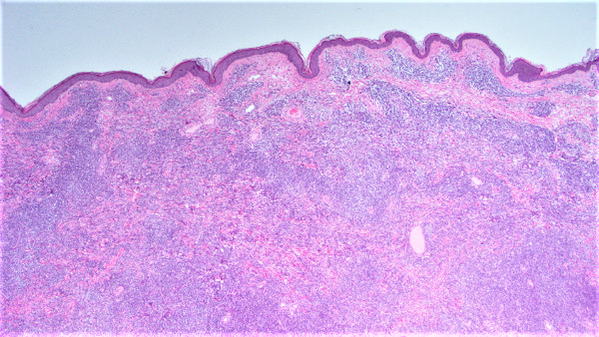
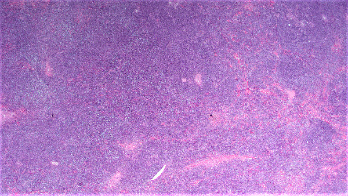
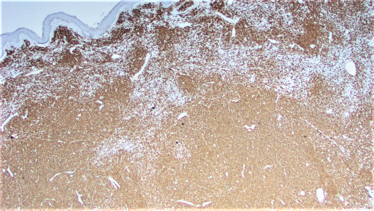
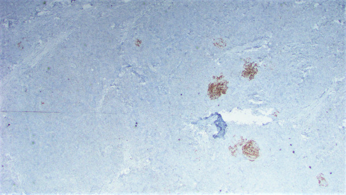
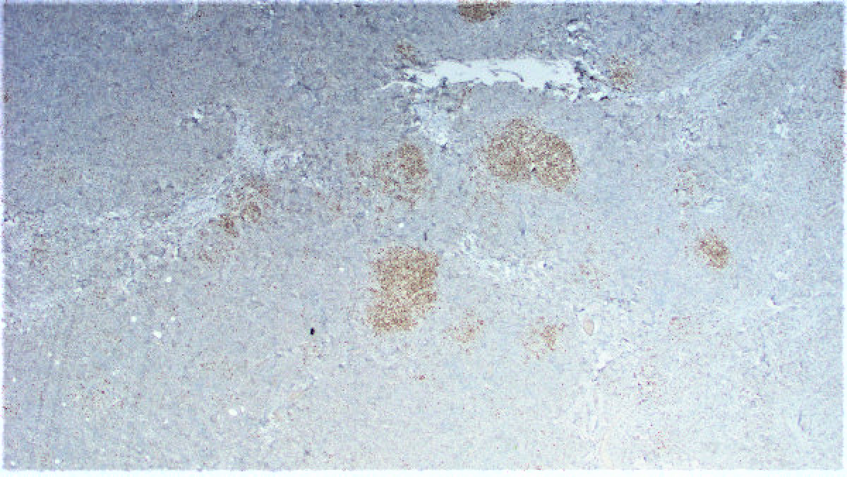
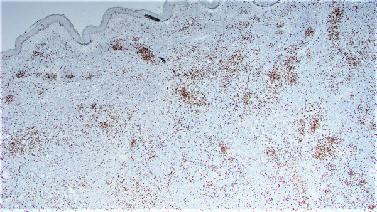
Three months ago the patient presented with a pink rash on the upper right arm. This was treated with topical steroids and the biopsy of that lesion revealed a reactive lymphoid proliferation. One month later the patient noticed a raised lesion on her upper right arm. An excisional biopsy of this lesion was performed. This biopsy revealed a vaguely nodular dermal infiltrate composed of a mixture of small cells with a high nuclear:cytoplasmic ratio and some larger cells with a plasmacytoid appearance (Figures 1 and 2). Immunohistochemical stains were performed and revealed both populations of cells to diffusely express CD20 (Figure 3) while negative for CD23 (Figure 4) and BCL-6 (Figure 5). There were scattered CD3 lymphocytes (Figure 6). Ki-67 was low (~20-30%), but elevated within residual germinal centers. Flow cytometry revealed lambda-restricted B cells, negative for CD5 and CD10.