Case 6: Upper GI Masses
Case 6: Upper GI Masses
By Aaron R. Huber, D.O.1; Gregory A. Gates, D.O.2; Tom C. DeRoche, M.D.3
Clinical History
A 75-year-old Caucasian male presented for elective esophagogastroduodenoscopy (EGD) for prolonged anemia and melena of uncertain etiology. The patient had recently been diagnosed with normochromic, normocytic anemia and melena requiring transfusion.
Imaging
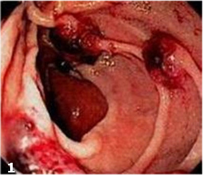
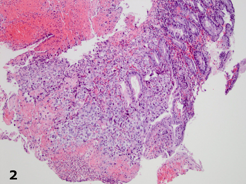
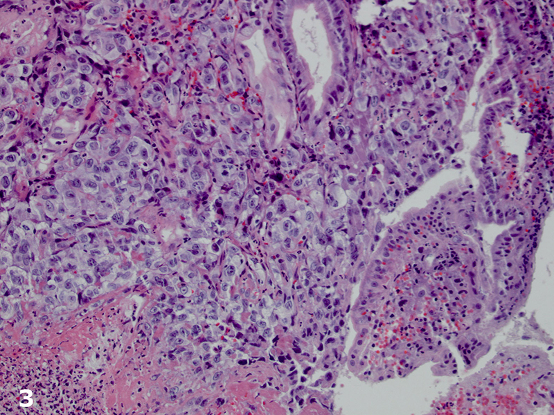
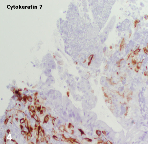
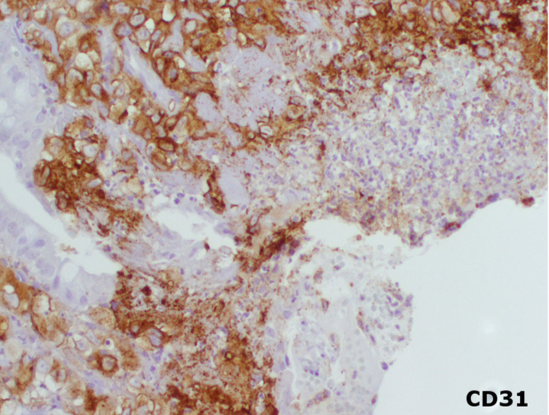
Imaging studies (computed tomography scan of the abdomen and chest radiograph) did not reveal a source for the bleeding or a mass lesion. The EGD demonstrated multiple red-brown polypoid masses within the stomach and duodenum (Figure 1).The biopsies, of both the stomach and duodenum, demonstrated large cells with amphophilic cytoplasm and irregular nuclei with vesicular chromatin and prominent nucleoli infiltrating the lamina propria (Figures 2 and 3). There was subtle vasoformation and, notably, no overlying dysplasia was identified. The neoplastic cells were positive for pan-cytokeratin, cytokeratin 7 (Figure 4), CD31 (Figure 5), and CD34. The neoplastic cells were negative for CDX2, TTF-1, S-100 protein, cytokeratin 20, cytokeratin 5/6, and calretinin.
Next: Diagnosis and Discussion
1. University of Rochester Medical Center, Rochester, NY 2. Naval Medical Center San Diego, San Diego, CA 3. Kaiser Permanente, Portland, Oregon