Case of the Month: Numerous Breast Lesions
Case Authors: Giby V. George, MD and Tatsiana Pukhalskaya, MD (PGY-4)
Clinical History
A middle-aged adult female presented with a firm breast mass. The lesion was not painful, nor accompanied by nipple discharge. In addition, palpation of the ipsilateral axilla revealed several enlarged lymph nodes.
Past Medical History
The patient reported to be overall very healthy. At the time of hospitalization she denied any medication except hormone replacement therapy.
Recent History
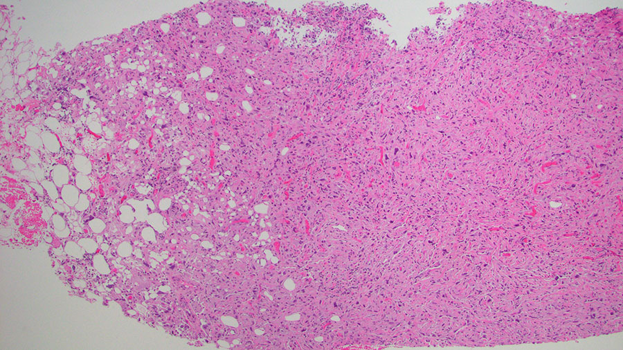
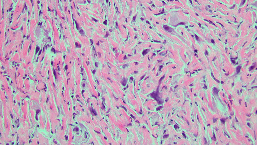
A MRI study revealed a main mass measuring up to 2.6 cm. There were numerous additional masses in bilateral breasts, with at least three displaying a highly suspicious predominantly washout pattern of enhancement. Core biopsy was performed with histology demonstrating pleomorphic spindled and epithelioid cells with abundant eosinophilic cytoplasm in a collagenous stroma (Figures 1, 2). An immunohistochemical workup showed tumor cells expression of GATA3, p63 and CD10. The tumor cells were negative for several cytokeratins (pan-CK, CK5, CAM5.2 and HMW) as well as e-cadherin, S100, SOX10, CD34, caldesmon, calponin, SMA and MSA.